"Purchase 20 mg isosuppra, acne 404 nuke book download."By: Joshua C Briscoe, MD - Medical Instructor in the Department of Psychiatry and Behavioral Sciences
- Medical Instructor in the Department of Medicine
 https://medicine.duke.edu/faculty/joshua-c-briscoe-md
Cheap 5mg isosuppra visaLoss of tactile discrimination and position and vibration sensation; taking pictures ache and paresthesias; Romberg signal. Intervertebral disk herniation 90% L4-S 1 1 0% C5-C7 Prolapse, herniation of the nucleus pulposus through faulty annulus fibrosus and into vertebral canal, impinging on spinal roots. Other clues: History of heavy lifting, optimistic leg-raise check, no aid with sitting. Involves enlargement of the central canal of the spinal wire, damaging fibers of the spinothalamic tract. Cranial Nerve Lesions Although most lesions are easy, some are regularly examined for pecu liarities (Table 6-2 1). Superior rectus I nferior indirect I nferior rectus Superior indirect Extraocular muscular tissues and their corresponding eye motions. Facial motion, style from anterior 2/3 of tongue, lacrimation, salivation (submandibular and sublingual glands), eyelid closing. Taste from posterior 1 /3 of tongue, swallowing, salivation (parotid gland), monitoring carotid physique and sinus chemo- and baroreceptors. Taste from epiglottic region, swallowing, palate elevation, speaking, thoracoabdominal viscera, monitoring aortic arch chemo- and baroreceptors. Thus, imaginative and prescient from the opposite intermediary cells and ship data down axons that kind the Ganglion cells of the retina: Receive input from the rods and cones through mediate shade imaginative and prescient. Commonly as a end result of mass-occupying lesions in the mind, extreme hypertension, or noncommu nicating hydrocephalus. The optic tract is formed from fibers from the ipsilateral temporal hemiretina and contralateral nasal hemiretina. The geniculocalcarine tract (visual radiation) pro jects via two divisions: the higher division and the decrease division (Meyer loop). The higher division passes through the parietal lobe, and Meyer loop passes by way of the temporal lobe. The posterior space of the visual cortex receives macular enter, or central vision, the intermediate space receives perimacular input, or peripheral vision, and the anterior area receives monocular input. Lesions at the numerous areas of the pathway create totally different visible defects (Table 6-22). The close to reflex and accommodation pathway permits pupils to constrict and focus on close to objects. Note that lesions proximal to the chiasm can be associated with monocular and ipsilateral defects, and lesions distal to the chiasm lead to contralateral and binocular defects. Bitemporal hemianopia, usually from an enlarging pituitary tumor in adults and a craniopharyngioma in children. Primary auditory cortex - Medial geniculate body Lateral lemniscus Nucleus lateral lemniscus Lateral lemniscus -. Sensorineural deafness: Caused by disease of the cochlea, cochlear nerve, or central auditory pathways. Rinne test: Place a vibrating tuning fork on the mastoid course of behind the ear until the patient can no longer hear the sound. Once the patient now not hears the sound, the tuning fork is held in entrance of the ear. Static labyrinth: Consists of the utricle and saccule and responds to lin ear acceleration of the head, together with gravity (head position). In these patients, the Rinne take a look at is regular as a result of the lesion causes nerve deafness. However, the Weber take a look at may be normal also if the nerve deafness affects both ears equally. Gustatory System lack of correct formation of the olfactory tract during growth. Hypothalamic neurons depend on the olfactory tract to migrate to thei r locations. J endrites: Mferent single or multiple extensions of the cell membrane that obtain indicators from other neurons or the setting of the neuron. Axons: Efferent extensions of the cell membrane that ship signals away from the cell physique to other neurons or finish organs. Neurons may have any number of dendrites and axons, which can be utilized for classification purposes: � Unipolar neurons have one dendrite or one axon. Pseudounipolar neurons have one course of that branches into a dendrite I and an axon. Neuronal axons contain each areas that are myelinated and those which are unmyelinated. Contain a excessive density of ion channels corresponding to voltage-gated Na+ channels that allow present to flow throughout the axon membrane. In the periphery, degradation and phagocytosis of the axon and myelin is followed by proliferation of Schwann cells. It is characterized by: � Disruption and dispersion of Nissl bodies � Rearrangement of the cytoskeleton with neuronal swelling � M arked accumulation of intermediate filaments Microglia: Phagocytes of mesodermal origin with irregular nuclei and little cytoplasm. They proliferate round injured nerve tissue and transform into large ameboid phagocytic cells in response to tissue harm. They help in axonal regen eration by creating a pathway for axon growth and secreting growth factors. The small spikes on the postsynaptic membrane characterize receptors for neurotransmitters. S ynaptic cleft: Site where exocytosed neurotransmitter molecules diffuse across to the postsynaptic membrane. Receptors: Bind the neurotransmitter and facilitate depolarization of the postsynaptic membrane by activating Na- channels. Dantrolene can be used to treat these circumstances (blocks the ryanodine receptors, inhibiting calcium-induced calcium release). Lambert-Eaton syndrome is an autoimmune disease in which antibodies assault presynaptic voltage-gated Ca 2 + channels of the neuromuscular junc tion (see Table 6-2 3). Sensory Corpuscles Neurons receive sensory alerts through the skin by way of specialized sensory organs: Meissner, Pacinian, Merkel, and Ruffini corpuscles, muscle spindles, and Golgi tendon organs. Other sensory organs embrace joint receptors, stretch receptors, baroreceptors, and hair cells of the inner ear (see Table 6- 1 7). Meissner and Merkel corpuscles: Mediate discrimination of nice spatial variations. Manifests with problem rising from a chair and weak point of huge muscular tissues especially within the morning. Golgi tendon organs (in collection with muscle fibers): Sense tension of ten dons and muscular tissues.
Buy 30 mg isosuppra with visaChapter 2 � Sinonasal and Craniofacial Region, Including Cranial Nerve V 77 Venolymphatic malformations may be classifed into three groups as advised by the World Health Organization: a. Lymphangioma simplex or capillary lymphangioma composed of thin-walled lymphatic spaces in regards to the measurement of capillaries that happen in the orbit, lip, cheek, tongue, gums, and f oor of the mouth the place the tight connecti ve tissue restricts the size ofindividual cystic areas b. Cavernous lymphangioma containing dilated lymphatic spaces intermixed with fbrous adventitia c. Cystic lymphangioma or c ystic hygroma composed of macrocystic lymphatic areas measuring from milli meters to se veral centimeters in diameter. Therefore, cystic hygromas are sometimes seen within the pos terior triangle of the neck. Syndromic associations of v enolymphatic malformations embrace Turner, Klinefelter, and Noonan syndromes. These lesions are often treated sur gically, b ut direct injection with sclerosing agents is a viable alternative for some patients. The goals are to relie ve useful prob lems, such as with airvay and feeding, while acquiring the very best cosmetic outcome. Islands of the malformation may be left behind pur posefully in order not to sacrif ce function. Subtotal resection is more probably within the lymphangioma or mixed v arieties ofvenolymphatic malformations than in cystic hygromas however mainly depend upon the placement relative to critical neu rovascular buildings. Reporting Responsibilities In basic, v ascular malformation is a main dif ferential diagnosis at the time of imaging, so no special communica tion is required. Any time a vascular malformation locations the airway in danger because of obstruction, communication with the referring remedy supplier and documentation ofthat communication is neces sary. If the malformation is complicated by inf ection, direct communication is also essential. Also, the orbital f oor defect persists and the medial rectus muscle is adherent to the realm of the defect, probably injured, a. Although there w as extensive medial antrostomy, the affected person e xperienced recurrent sinus signs perhaps as a result of the uncinectomy was incomplete. Anticipation of ana tomic v ariants, similar to a more medial than usual position of the medial orbital wall on the preoperative research, would possibly assist to avoid the extraordinarily unfortunate orbital entry and eye issues that occurred in this case. Reporting Responsibilities In this case, the patient has suf fered a signif cant complica tion that has long-term implications with re gard to ho w nor mal ocular motility could be restored. The most posterior ethmoid cell can pneumatize f ar lat erally and superiorly to the sphenoid sinus. When such pneumatization happens, the cells are referred to as sphe noethmoid cells or Onodi cells. Recognition of this ana tomic v ariant might help the sur geon a void injuring the carotid artery and the optic nerv. The scalp abscess reveals f ndings suggesting comparatively thick and purulent material (arrow). There is proof of epi dural abscess (arrowhead) displacing the superior sagit tal sinus. The superior sagittal sinus is displaced (arrowheads) by the epidural abscess (arrow). P atients with a history of aller gy, occupational or vasomotor rhinitis, and anatomic obstruction corresponding to septal deviation or nasal polyps have a tendency to infections. The condition can also be extra frequent in sufferers with immune def ciency and ciliary motility issues. There was bone erosion of the anterior table of proper fron tal sinus with abscess formation in the deep scalp. However, no direct connection of the intracranial subdural emphyema and the contaminated sinus could be established. The unfold to the epidural area may be direct by way of adjoining bone or through transdural veins-the latter occurring on this patient. Other complications that may be seen with acute sinusitis are pre- and postseptal orbital cellulitis, subperiosteal and orbital abscess formation, cavernous and other dural venous sinus thrombosis, subdural empyema, meningitis, and brain abscesses. Reporting Responsibilities Direct communication is strongly really helpful for all instances the place imaging is finished for the suspicion of issues of an acute sinusitis. The impor tance of imaging is to e valuate the e xtent of disease, detect clinically unsuspected complications, and determine attainable structural etiology which will have predisposed the patient for such an episode. The coordinated action of the cilia of the columnar epi thelial cell mo ve the sinus contents to ward the pure sinus ostia. Disruption of the ciliary function leads to accumulation of sinus secretions and doubtlessly infec tious brokers within the sinus. Chapter 2 � Sinonasal and Craniofacial Region, Including Cranial Nerve V 83 Various conditions can affect the mucociliary function. This includes excessive airf ow and chilly air, toxins produced by microorganisms, environmental mediators of the inf ammatory response, mechanical f actors that impede transport of the mucosal blanl<et, major ciliary dyskine sia and secondary ciliary dysfunction from persistent infec tions, and secondhand smoke publicity. When the natural sinus ostia become obstructed, nor mal mucus drainage is impeded. The obstructed sinus setting turns into hypoxic and causes ciliary dys perform and modifications in mucus manufacturing, all of this fin. It is this fundamental situation that creates a possible for the orbital and intracranial compli cations. It lik ely contrib utes to the pathophysiology of bone erosion and direct unfold of disease past the bony sinus limits as nicely. Relieving this stress by draining the causati ve sinus is a technique aimed at the prevention and treatment for such compli cations. Such drainage also promotes the restoration of normal sinonasal mucociliary drainage by reversing a variety of the parts causing that dysfunction. She has historical past of seasonal allergy symptoms and was used to recurrent and persistent episodes of sinusitis, however this headache has been bothering her extra. The contents seem dense and likely desiccated with a central area ofhigher density (arr ow). The sinus contents appear of some what greater signal intensity than f uid (white arrow). The edematous mucosa inside the sinus once more remains relatively thin (arrows) with the polypoid mucosal thick ening in the sphenoeth moidal recess and around the sphenoid ostium inflicting the first obstruction somewhat thicker (black arrow). These circumstances are most typically encountered in patients with persistent rhinosinusitis and particularly those with nasal polyposis. A prior history of either trauma or earlier sinus or f acial surgery can also be encoun ti. There must also be confrimatory endoscopic infammation and proof of rhinosinusitis on imaging. P atients with a historical past of allergy, occupational or v asomotor rhinitis, nasal polyps, or anatomic obstruction such as septal de viation and concha b ullosa have a tendency to continual infec tions. Rhinosinusitis is extra frequent in immunodef cient sufferers, those with ciliary motility problems similar to Karta gener syndrome, and those with mucous blank et issues corresponding to sufferers with cystic f brosis. Most chronic sinusitis is now thought to be noninfectious in etiology, though infec tion could have incited the method.
Purchase 20 mg isosuppraAll opioid receptors are linked through G proteins and inhibi tion of adenylate cyclase. Opioid antagonists similar to naloxone and naltrexone could be given for overdose, as these drugs Summary of mediators derived from arachidonic acid and their actions, and websites of action for anti-inflammatory medicine. Liver damage may be prevented if N-acetylcysteine or methionine are given, as they regenerate glutathione. In the third gesta tional week, following gastrulation, the neural tube forms, and neural crest cells emerge and migrate, starting the exactly controlled growth of the central and peripheral nervous methods, respectively. E ctodermal cells detach from the epiblast, the floor layer of the embryo, invaginate inward into a groove often known as the primitive streak, and kind the mesoderm and endoderm. Mesodermal cells within the primitive streak then migrate towards the pinnacle until blocked by the fused buccopharyngeal membrane at the primitive node (the most rostral a half of primitive streak). In parallel, prenotochordal cells also invaginate and move rostrally, kind ing a line often known as the notochord from the primitive node to the pre chordal plate. Neurulation the primitive streak regresses and disappears, dragging the notochord toward the buccopharyngeal membrane. The notochord becomes the nucleus pulposus, which lies inside the vertebral column in the grownup. Herniation of the nucleus pulposus by way of the annulus fibrosus may end in spinal root impingement and ache. The notochord induces the overlying region of the ectoderm to type the � � the neural plate begins to invaginate alongside the longitudinal axis and forms the neural groove. The open neural tube then closes, starting within the middle (at the center of the longer term body) and progressing caudally and rostrally. Invaginating mesoderm cells (prenotochordal cells) detach from the epiblast and migrate along the longitudinal axis to kind the notochord. Failure of rostral neuropore closure leads to anencephaly, a situation characterised by the absence of the scalp, cranium, and huge portions of the cortex. Elevated a-fetoprotein in maternal serum or amni otic fluid is often suggestive of fetal neural tube defects. Failure of vertebral arches to shut with herniation of meninges however not spinal twine. Failure of vertebral arches to shut with herniation of each meninges and spinal cord. Herniated lumbosacral sac and, depending on the location, paralysis and loss of deep tendon reflexes and sensation in the lower extremities as well as incontinence. Meningocele Meningomyelocele (myelo = neurons/cord) Meningoencephalocele (encephala = brain) Herniation of meninges and mind. In the mind stem and spinal wire, the dorsal alar plate, the ventral basal plate, and the intervening sulcus limitans develop inside the central canal of the neural tube. Ectoderm at the edges of the neural folds is induced by the neural tube to type neuroepi thelia. The most typical issues of neural crest cell migration embrace these listed in Table 6-2. The neural crest cells migrate peripherally from the neural tube to become the peripheral nervous system and different important structures. The 5 secondary vesicles are: � � � � Telencephalon (derived from the prosencephalon). Type I could not show neurologic signs until adolescence or adult life and should embrace cerebellar ataxia, obstructive hydrocephalus, brain stem compression, and syringomyelia. Death is normally as a result of cranial nerve and brain stem dysfunction leading to respiratory failure. Arnold-Chiari syndrome is rather more widespread and includes a "falling through" of the cerebellum. Dandy-Walker syndrome is an agenesis of the cerebellum and a enlargement of the posterior fossa. The elevated intracranial stress pushes the bones apart, enlarging the cranium and head circumference. Growth of the cranium is determined by development of the mind, so the top circumference is decreased sec ondary to the underlying defect in mind improvement. This could additionally be due to genetic causes, prenatal an infection, or publicity to teratogens (eg, toxoplasmo sis, alcohol, radiation). This could be seen in severe fetal alcohol syndrome and Patau syndrome (trisomy 1 3). Severe instances, nevertheless, can present with midline structures, and a single ventricle. The is dependent upon the extent to which constructions are commonest reason for menta l retardation. I t is characterized by microcephaly, congenital heart illness, abnormal facies, and, in severe instances, holoprosencephaly. In delicate cases, individuals could lead a normal life; in more extreme circumstances, the situation might result in mental retardation and early demise. S ylvian fissure = oblique groove that divides the temporal lobe from the parietal and frontal lobes. Central sulcus = major sulcus that runs coronally, dividing the frontal lobe from the parietal lobe. Autosomal dominant polycystic kidney illness, hypertension, smoking, blacks, i age. Pia mater: Delicate and highly vascular; intently adheres to the floor of the brain and spinal twine. It is a comparatively slim area over the floor of the cerebral hemispheres, however it becomes a lot wider on the base of the brain. Subdural space: Lies between the arachnoid and the dura and is traversed by bridging veins within the brain. Epidural house: Potential house exterior the dura that incorporates meningeal arteries. In the spinal cord, it contains fatty areolar tissue, lymphatics, and -,� enous plexuses. Bleeding within the meningeal spaces manifests in a different way depending on the supply and location of the bleed. Usually as a result of blunt head trauma leading to rupture of the center menin geal artery. Imaging shows a biconvex assortment of blood bordered by suture strains with lens-shaped enhancement and clean borders. Often accompanied by intracranial bleeds or contusions and presents as decline in psychological standing over days to weeks. Subdural hemorrhage is seen most often within the elderly as cortical atrophy puts rising tension on bridging veins but can be seen in alcoholics and shaken infants.
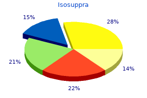
Trusted 10mg isosuppraAppears much like leiomyomas however diagnostic features embrace pleomor phic spindle cells with relatively frequent mitoses. Polycystic Ovary Syndrome Characterized by chronic anovulation, hirsutism, obesity, and enlarged poly cystic ovaries. Although the ladies are insulin resistant, the ovaries are stimulated by the insulin to secrete extra portions of androgens. Follicle progress is stunted, with follicles reaching a maxi mal dimension of 2-9 mm in diameter. They are additionally at elevated danger for endometrial hyperplasia and automotive cinoma as a result of extended unopposed estrogen exposure. Sonogram exhibiting enlarged ovaries, with a number of (> 1 2) small cysts (2-9 mm in diameter) in a "string of pearls" configuration. Other endocrinopathies should be ruled out, such as hypothyroidism, hyper prolactinemia, or late-onset congenital adrenal hyperplasia. However, the corpus luteum can typically accumulate fluid, thus becoming a corpus luteum cyst. It can develop up to 6 em in diameter and has a possible to rup ture, which consequently may cause ovarian torsion. There is an affiliation of corpus luteum cysts with using ovulation-inducing medicine corresponding to clomiphene citrate. Theca Lutein Cyst Lined with theca interna cells, theca lutein cysts are usually bilateral and often regress spontaneously. The cysts are associated with molar being pregnant, choriocarcinoma, twin pregnancy, Rh isoim munization, and ovulation-inducing brokers such as clomiphene citrate. Neoplasms Asymptomatic till rising tumor turns into giant sufficient to produce symp toms of belly distention or fullness, or a dragging sensation as a end result of mass effect. A tumor mass also predisposes to ovarian torsion, inflicting intermittent intense and sharp ache. Constitutional signs of fever, chills, and uninten tional weight reduction may also be present. Often present at early levels, in contrast to epithelial ovarian tumors, that are sluggish growing and often present at late stages. Sonogram may reveal adnexal mass measuring > 2 em with cystic or solid elements. Histologically, dysgerminomas exhibit massive, spherical cells with clear cytoplasm and enormous nuclei with distinguished nucleoli. Blood vessels with most cancers cells resembling primitive glomeruli (Schiller-Duval bodies). Appears as an ill-defined invasive mass containing foci of hemorrhage and necrosis. The cells are giant and primitive wanting, with basophilic cytoplasm, indis tinct cell borders, and enormous nuclei with distinguished nucleoli. They comprise differentiated somatic cells from a number of germ layers (ectoderm, mesoderm, and endoderm). Teratomas are agency plenty that on cut floor often contain cysts and recognizable areas of cartilage. Contains well-differentiated bone, cartilage, hair, muscle, and/or thy roid follicles. It usually seems at the fifth or sixth decade of life and accounts for 90% of all ovarian cancer. The use of oral contraceptive drugs has been documented to help stop ovarian most cancers. Characterized by ingrowths of papillary and glandular structures with stromal invasion. Characterized by a quantity of loculi lined with mucin-secreting epithe lium and stromal invasion. Pseudomyxoma peritonei is a potential complication and involves tumor production of mucus in the stomach cavity. If not handled, mucin will accumulate to such an extent that vital structures in the abdomen are compressed. Characterized by comparable adenomatous sample seen in endometrial carci noma of the uterus. Endometrioid carcinoma of the ovary correlates with concurrent lesions in the endometrium. Often current with stomach distention, pelvic or stomach ache, and irregular vaginal bleeding. In reproductive age group, associated with menstrual irregularities, endo metrial carcinoma (5 %), and endometrial hyperplasia (5 zero %). Grossly, smooth with lobulated surfaces; measurement can range from few millime ters to 20 em. Cells often organize themselves around a central cavity like a primordial follicle (Cali-Exner bodies). Because of the kind of hormone produced, generally presents with indicators of virilization corresponding to amenorrhea, breast atrophy, pimples, hirsutism, deepen ing of the voice, and receding hairline. When the Nitabuch membrane is deficient, the trophoblastic tissue attaches on to the myometrium. Incomplete separation of the placenta during supply results in profuse hemorrhage. The commonest loca tion for ectopic being pregnant is the ampulla of the Fallopian tube. The price of ectopic being pregnant is 2% of all pregnancies in the United States, with African Americans at elevated threat. On examination, abdominal tenderness and cervi cal motion tenderness could also be present, and an adnexal mass may be palpated. Amniotic fluid is produced by the fetus, aids in normal development and develop ment, and helps defend the fetus. Too a lot (polyhydramnios) or too little (oligohydramnios) amniotic fluid can lead to abnormalities within the developing fetus. Associated with fetal intestinal atresia, esophageal/duodenal atresia, anen cephaly, maternal diabetes, neural tube defects, and multiple gestations. This leads to vascular endothelial harm, vasoconstriction, hypertension, renal glomerular endothelial cell harm, and coagulation abnormalities. Intense vasospasm is induced by the discharge of varied mediators like endothe lin and thromboxane A2. The diploma of preeclampsia depends on the extent of trophoblastic invasion by the placenta. During labor and supply, magnesium sulfate might forestall extreme preeclampsia and eclamp sia.
Discount 40mg isosuppraCompression with axonal damage and/or demyelination ofthe nene will interf ere with its perform, inflicting hypere xcitability that results in hemif acial spasm or diminished operate inflicting facial weak spot. The facial nerve canal will usually seem to be focally expanded on the web site of origin. Spread btyond the racial canal is most typical at the f rst genu the place the malformations unfold within bone alongside the course of the larger petrosal nerve. Use ofsuch knowledge related to bone and calcif cation may not dif ferentiate hemangioma from the menin giomas that occur in this region. Heman giomas could additionally be handled by observ ation solely, depending on whether the f acial nerv e weak point is progressi ve; nonetheless, they typically happen in youthful inditiduals, will progressively enlarge, and shall be remo ved. In the era ofmodem imaging, many ofthese lesions are dis covered at a time when the y could also be treated by stereotactic radiosurgery with out histologic conf rmation. Radiotherap y ought to be delayed until the probability ofa nerve sheath tumor is established in small (3- to 5-mm) lesions since those would possibly truly be due to inf ammatory enhancement ofthe nerv e and the improve ment might resolv e without specif c therap y. Such f ndings may also require further testing such as L yme titers and syphilis screening before radiotherapy is initiated. What are some related caveats in making a medical deci sion to deal with small facial nerve lesions Reporting Responsibilities In slowly progressive disease or in sufferers with only hemif a nerve and perineural enhancement may also progress in a way extra in maintaining with perineural spread ofcarcinoma or neurotropic lymphoma and in the end lead to a dif f erent prognosis and treatment plan. What the Treating Physician Needs to Know � Does the imaging demonstrate a structural lesion from the seventh nerve nucleus to its peripheral site of innerv ation with a high degree of confdence Even at that point, persistent enhancement should embrace a consideration ofinf ammatory etiologies ofa extra continual nature than viral infammation. Reporting Res ponsibilities Routine reporting often suff ces except the lesion is one that means an aggressive illness course of corresponding to an infec tion or meningeal carcinomatosis. In very small (3- to 5-mm) enhancing lesions, one should take nice care to counsel that these could additionally be focal inf am by the f acial nerve presentation of this disease when that region of the central nervous system is involved. A proper vary of attainable inf ammatory circumstances ought to be talked about within the report to stimulate an acceptable workup. The risk of Lyme illness, perhaps e ven in nonendemic areas, should always be included within the reported differential analysis. Imaging of those tumors requires the very best spatial reso lution, especially for small intracanalicular schw annomas. The most essential imaging differ ential is a meningioma, which generally has a dural tail on postcontrast TlW sequence. Lar ger tumors normally turn into symptomatic because of mass impact on the brainstem and cerebellum resulting in ipsilateral upper and lo wer extremity dysfunction, ataxia mm sectin thickness post Questions for Further Thought 1. Reporting Responsibilities Routine reporting usually suff ces except the lesion is one that suggests an aggressi to ve illness process corresponding to an infection or meningeal carcinomatosis. In lar ger lesions that compress the brainstem and/ or trigger hydrocephalus, the need for direct communication might rise to an urgent and even emergent level. Initially, the affected person may be treated medically for symp tomatic relief As the tumor is entirely intracanalicular. Because ofthe unkno the goal ofstereotactic radiosurgery is to eliminate or arrest tumor wn long-term ef fects, it ge tumors with a contraindica will not be used in younger sufferers. Lar brainstem or cerebellar compression are tion for radiosur gery as a outcome of posttreatment edema might improve compression. It should be accomplished at no larger than 2-year interv als until the expansion fee is nicely established on serial exams. Failure to acknowledge the true nature of this mass w ould have led to an unnecessary craniotomy and a danger of disastrous bleeding. Also, a potential misadventure that might outcome from the lesion not being acknowledged as highly vascular until the time of swgery could be avoided. The finest treatment option could be endovascular embo lization of the feeding v essels. Thus, the f ndings of early labyrinthitis ossif cans within the second tum of the cochlea had been unrecognized preoperatively, probably as a end result of improper window and stage setting used while reviewing images. Are there an y abnormalities alongside the whole auditory pathway which may contraindicate or alter the prognosis of treatment by cochlear implantation The sur geon needs to be told in regards to the patenc y ofthe cochlea and any cochlear and vestibular anomalies or bone dysplasias in addition to an y retrocochlear disease. Are there any indicators ofcomplications which may trigger mal operate, or can dysfunction be anticipated by alterations in frequency mapping Imaging outcomes could alter the choice ofside ofimplantation or recommend the extra acceptable electrode selection. Compressive neuropathy from Eagle syndrome (gross elongation, enlage ment, and calcif cation of the stylohyoid ligament) could cause referred otalgia. The lesions in the abo ve se gments mostly in volve multiple lower cranial nerves. Ifthere is an altemati ve explanation for the symptoms or signs ofthe glossopharyngeal neuropathy af finish organ ofinnervation fecting the � the pyriform sinus. Progressive, presumably referred, otalgia should be seen with a really high index ofsuspicion even in light of unfavorable upper aerodigestive tract endoscopy, espe cially within the smoking/drinking population. Several branches supply sensation to the middle ear and bon y eustachian tube, the posterior oropharynx and taste bud, sensation and taste to the posterior third ofthe tongue. Hence, imaging is concentrated on all of these areas when sufferers current with "referred otalgia. Degree ofconf dence ofa ne gative research e xcluding sig nifcant causative pathology � Answer 1. Patients with glossopharyngeal neuropathy and referred mclude temporal bone and posterior skull base recon structions with the same technique used to study main temporal bone issues and high-detail pictures from the posterior fossa to the bottom ofthe p yriform sinus. For this purpose, no submucosal mass ofthe pharynx must be biopsied-whether or not associated with signs or signs possibly as a result of a lower cranial neuropathy with out prior imaging. Ifthere is a structural trigger for the neuropathy Is more than one nerve doubtlessly involved From the skull base to the carotid bifurcation, the patholo gies embody paraganglioma, meningioma and benign v agal origin schw annoma, nasopharyngeal carcinoma (due to direct unfold or retropharyngeal adenopathy), and untreated cervical nodal metastasis as nicely as recurrent metastatic nodal neck disease. Below the bifurcation ofthe carotid supracllzlicular lesions and much like lymphoma could cause v agal neuropathy. However, the more frequent lesion at his ltYel ofthe neck is both major thyroid cancer (as on this patient) or metastatic lymph nodes from thyroid cancer, esophageal most cancers, or tracheal adenoid cystic most cancers infltrating the recurrent laryngeal nerve within the tracheoesophageal groove. Thoracic pathologies that may cause v agal nerv e palsy include those of the aortic arch similar to an aneurysm and/or dissection and malignant mediastinal lymphadenopathy. The imaging st udies then primarily become an e valuation of a vagal and/or recurrent laryngeal nene neuropathy. Ifthe only different defcit is ofthe cervical sympathetics (Homer syndrome), then the lesion could lie between the skull base and thoracic inlet and other accompanying symptoms such as brachia! Ifthe only symptom is hoarseness and the only signal is an atrophic, paralyzed, or paretic tr ue wire, a cause is most incessantly not found by imaging; ho. The vocal wire paresis could be the solely sign ofconditions as di verse as v agal cistemal segment compression due to a posterior fossa meningioma, a delicate mass in the thyroid gland or mediastinum causing compression or inf ltration ofthe recurrent laryngeal nerv e in the tracheoesophageal groove or aortopulmonary windo. Other vascular pathologies similar to carotid or aortic dissection, life threatening intracranial abnormality similar to hydrocephalus, or brainstem compression need to be communicated imme diately. Degree of conf dence of a ne gative research e xcluding � signifcant causative pathology Answer 1. Sometimes no structural lesion is identif ed in a affected person with a specif c cranial neuropathy. Imaging research in sufferers with indicators and signs of a possible cranial neuropathy similar to otalgia or v ocal cord paralysis will as a rule be adverse. The most important thing to bear in mind is to mak e sure that the research is of fine high quality and has been carried out with the right pro tocol for the suspected pathology. If the signs become w orse or an additional cranial neuropathy declares itself or turns into suspect, the patient ought to be followed with one other imaging exam.
Order isosuppra 5mg on-lineThe e xercise is to acknowledge the dif ference between the tw o spaces and the diagnostic implications of correct identif cation of the space of origin. When because of inf ammatory illness, the scientific situation may well dictate the correct interpretation of retropharyngeal adenopathy. In the occasion of retropharyngeal adenopathies and an unknown major cancer, cautious inspection of the pharynx could reveal a clinically occult, submucosal major lesion. Reporting Responsibilities When these research are carried out to waluate a affected person with a pain sample that could be related to the spine and/or pharynx, the discovery of such a mass should provoke direct communica tion. If the method has an y potential to cause spinal cord compression, v erbal contact should be escalated to ur gent or emergent. Timely analysis and correct remedy are crucial in pre venting sequelae such as airw ay obstruction, mediastinitis, carotid artery aneurysm, and ca vemous sinus thrombosis. True retropharyngeal abscess, apart from that because of pen etrating trauma, iatrogenic trauma, or some other explanation for pharyngeal perforation, is unusual. Common clinical shows in adults embrace fever, odynophagia, and dysphagia. Occasionally, these pus-containing nodes turn into so lar ge that the y represent an abscess physiologically. Infection or abscesses could unfold from different suprahyoid areas and could also be related to skull base osteomyelitis. The airway should be controlled and the patient handled with intravenous antibiotics. Sur gical drainage of suppurati ve retropharyngeal lymph nodes could also be required for these sufferers with persistent or enlarg ing nodes which may be unresponsive to aggressive antibiotic ther apy and maybe those that exceed three cm in most axial What the Treating Physician Needs to Know � Likely diagnosis and de analysis gree of conf dence in that � Whether the patient is in any imminent danger Full extent of the infectious process Relationship of the an infection or inf surrounding anatomy ammation to critical � � brief axis dimension and/or seem lik ely to rupture. Of course, immediate surgical drainage might turn into necessary in patients with airway compromise. In this case, there ws direct communication with each the pediatric and otolaryngology companies in order that both understood this disease would likely be managed with intravenous antibiotics and never require surgical drainage. There is an inside air-fuid level (arrow) and a thick minimally enhancing and relatively well-demarcated wall. One have to be f amiliar with the complete range of pathology attainable as a end result of patients with neck plenty of unsure etiology generally present confusing scientific problems and/or physical fndings that can be greatly simpli f ed by good-quality, well-interpreted pictures. This makes branchial and thymic equipment cysts unlikely; nevertheless, in this developmental cat egory, it could be affordable to think about a speaking foregut duplication cyst. The air-f uid stage suggests a connec tion with the aerodigesti ve tract or gas-forming micro organism. Many neck mass cases require direct communication with referring doc tors to resolve on a plan of action, especially if imaging directed biopsy is important. In the low neck, the method spreads throughout the visceral compartment and into the lateral compartment (arrows). Infectious illness originating from the cervical spine must be differentiated early in the diagnostic process from that orig inating due to pharyngeal illness to a void a probably cata strophic neurologic event involving the cervical spinal twine. The process may be supplemented by imaging directed aspiration and/or tissue sampling, which may be very secure. Infections that arise in a diabetic or in any other case immunocompromised patient usu ally require some direct communication. What the Treating Physician Needs to Know � Likely diagnosis and degree of conf dence in that diagnosis Full extent of the infectious process Relationship of the infection or inf surrounding anatomy ammation to critical � � � If any extra data must be collected with imaging assis tance Posttreatment imaging: Suspicion for recurrent or persis � tent infection versus expected posttreatment modifications Determine whether the method includes single or a number of areas. If single, which house is involved, primarily based on making obser vations with re gard to v ectors of structural displacement and spread of the inf ammatory course of Occasionally, those pus-containing nodes become so lar ge that the y constitute an abscess physiologically. The origin is lik ely from v arious spinal nerve roots and probably the sympathetic ple xus (arrows). The extension into the neural foramen (arrow head) on the left clearly identifes no much less than one segmental cervical nerve root of origin. Transspatial processes are, re gardless of area of origin, lots that arise primarily within the deep spaces of the neck. For aggressive pathologies similar to cancers or diskitis and vertebral osteomyelitis, transspatial unfold is a simple morphologic unfold pattern to understand. It can be is a:Birly intuitive pattern to perceive in d(Yelop mental abnormalities that have an effect on these areas since enolym phatic malformations simply observe vessels as they develop. Identify associated f ndings corresponding to lymphadenopathy epidural illness, diskitis, or tendon and ligament calcif cation. This favors the analysis ofneurogenic or nene sheath tumor corresponding to pled kind neurofbroma. Nerve sheath tumors are clearly the kind of lesion that can show transcompartmental unfold is as follows: � Determine whether the process entails single or a number of areas. Chapter 5 � lnfrahyoid Neck and Cervicothoracic Junction (Thoracic Inlet) 223 since they follow nerves by way of anatomic gaps and throughout boundaries between these compartments. What the Treating Physician Needs to Know � Likely analysis and degree of conf dence in that diagnosis Full extent of the mass Relationship of the mass to critical surrounding anatomy If any extra knowledge must be collected with imaging assis tance Posttreatment ima ging: Suspicion for tumor recurrence Question for Further Thought 1. Many of those instances require direct communication with referring docs to decide on a course of action, especially if imaging-directed biopsy is necessary. This communication might turn out to be urgent or emergent if the an infection wolves or could herald disease within the epidural space which may threaten the spinal cord. The general mechanism of these de velopmental errors is incomplete obliteration of the branchial equipment lea ving Their embryologic ori arious medical presenta arious c ysts, sinuses, and gin e xplains the di verse appearance of those anomalies on tions. For the security of patients, the entity "most cancers arising in a branchial cleft c yst" have to be thought of an impossibility and the cystic mass presumed to be a nodal metastasis from most cancers of the pharynx or larynx, pores and skin (ifparotid region), or thyroid ifin the low neck. Reporting Responsibilities Branchial apparatus developmental anomalies are generally chronic entities that may be reported routinely. Classif cation is completed primarily to anticipate anatomic relationships that may be important treatment concerns, such as the course and level of pharyngeal communication of a fstula or sinus tract that has turn out to be difficult by infection so that it may be treated utterly. Cysts, as on this case, will present as a neck mass, often because they turn into secondarily infected. The mass is often in the usually will end up to be because of a metastatic node, and a dili gent search of the pictures for a potential primary tumor and direct communication to the referring supplier are essential. Communication might include identif cation of the primary supply or a suggestion for needle aspiration/biopsy of the mass. What the Treating Physician Needs to Know � Are the f ndings because of a branchial apparatus anomaly or presumably one other condition What is the arch of lik ely origin to anticipate a inside fstula or sinus tract location If a cyst, its full extent relative to surrounding anatomy Is there an associated sinus or f stula Is the f nding associated with glandular ectopias or pathol ogy arising in ectopic glandular tissue Cysts most typically relate to the anterior triangle b ut may be lateral to the carotid sheath in order that those projecting extra to ward the posterior triangle are more doubtless related to the third cleft. A historical past of intermittent modifications in measurement or inf amma� � � � tion will counsel a branchial cleft c yst as a attainable etiol ogy, particularly in youngsters and young adults. Third equipment cysts could present as recurrent inf ection in the re gion of the thyroid gland (the e xact branchial equipment origin of those remains somewhat controversial). The younger the affected person, the more a de velopmental prob lem might be believable, but great care have to be taken in adults. A predominantly c ystic mass could additionally be a nodal metastasis, and this risk have to be defnitively excluded, especially in this case where the patient has a previous historical past of alcohol and nicotine abuse. The mass is centered primarily in the ante rior cervical triangle and is bilateral.
Buy isosuppra 20mg lowest priceThe lesion has no unique options and it was biopsy that exposed the diagnosis ofmalignant fbrous histiocytoma. The mechanisms and potential routes oftrans spatial unfold of these aggressive pathologies such as lymphoma, metastases and sarcomas are by direct invasion and/or perineural along the cervical nerve roots probably to the epidural house and cervical spinal twine. Describe the wctors ofstructural displacement and spread of a mass centered within the paravertebral area. Reporting Responsibilities Malignant tumors ofthe posterior compartment may run a spectrum from people who pose no quick consequence to those that may result in fast neurological deterioration due to spinal wire in volvement that may not be predictable from the f ndings. Direct, ur gent communication with the referring remedy provider is important within the latter circumstances and all the time at the time of initial suspicion ofa malignant mass. Chapter 5 � lnfrahyoid Neck and Cervicothoracic Junction (Thoracic Inlet) 243 the primary components of the report ought to embrace: � Answer volved 1. Superiorly, the process could strategy the posterior cranium the positioning of origin and all spaces/compartments in base above from the thoracic inlet and mediastinum belcw to the skull Whether the spinal cord is potentially involved Risk of the mass being malignant base within the neighborhood of the upper cervical spine or fora males magnum. Inferiorly, the mass might track to the supraclavicular fossa and into the upper back. Medially, the process will occup y the rnidline or a paramedian place; it might in volve the backbone and neural components. Cervicothoracic junction plenty may also be related to other branchial cleft or pouch dysgenesis, a prime e xample being the contaminated p yriform sinus tract or third branchial cleft c yst and associated tracts that the majority fre quently present as an infection within the lo w neck around the thyroid gland. Duplication c ysts of the fore intestine and neuroenteric c ysts could also be a uncommon explanation for a thoracic inlet mass. More com monly, cysts and "celes" related to the pharynx, such as wi ous di verticuli, will happen at websites of natural weaknesses at developmental boundaries between muscular tissues or along pen etrating neurovascular bundles. Rests of germ cell layer tissue might produce epidermoid, dermoid, and teratomatous plenty in the lo w neck and tho racic inlet. Rests of salivary gland tissue are also left within the deep areas of the top and neck throughout de velopment. These could gi ve rise to each benign and malignant sali vary epithelial tumors at nearly ary neck level, however these tumors are uncommon at this stage. Neurologic de velopmental abnormalities similar to those arising from neural crest remnants and dysraphisms can current as a neck mass. The price of incidence and patient actors similar to age should be taken under consideration. An etiologic method based mostly on developmental origin must be the primary diagnostic strategy to pediatric sufferers and young adults as quickly as the thyroid is excluded as an etiology. Thymic migration follows the trail of the third branchial equipment from the extent of the carotid bifurcation within the lat eral compartment to the midline in the lcw neck, terminating within the mediastinum so that various iterations of thymic rem nants may current within the low neck and thoracic inlet. Devel opmental cysts are mainly related to the thymus and as such are inclined to be in the midline, then veering to the left. Venolymphatic malformations are relatively common and essentially the most ubiquitous of the transcompartmental benign plenty encountered in the low neck and thoracic inlet. A specifc surgical strategy will rely heavily on the extent and origin of the mass as determined from imaging. Decisions about how a lesion is approached depend upon the site of origin and its relationship to the supracla vicu lar fossa, brachia! The rela tionship of the mass to the brachiocephalic v essels, par ticularly the carotid and vertebral arteries, could additionally be pivotal in choice making. The report ought to include precise element about the full extent of the mass and relationship to critical surrounding anatomic structures that could be the origin of the lesion and/or affected by swgical or other remedy. In general, essentially the most crucial of these relationships are ho w the mass relates to the trachea, brachiocephalic v essels, brachia! It is very necessary to talk eff ectively and promptly ifthere is a secondary an infection and in these instances to anticipate whether there may be a complicating sinus or fstula. Flow-sensitive gradient echo image shows that the right subclavian is actually clotted; that f nding is further conf rmed on the maximum projection inten sity image in. In this specific case, mechanical irritation and compres the scapula posteriorly and laterally; the third and most dis tal area of attainable constriction is the subcoracoid space beneath the coracoid course of lying deep to the pectoralis minor tendon. Reporting could also be extra routine if a benign etiology or continual vascular condition is identif ed. What the Treating Physician Needs to Know sion resulted in inf ammation that also af fected the subcla vian artery w all. Supposedly the mechanical compression, the inf ammation, and presumably the harm to the arterial wall then resulted in an arterial occlusion. Reporting Responsibilities Compressive thoracic outlet situations may be a signif cant acute menace to the patient when acute or subacute arterial occlusion is feasible. Such circumstances require direct, instant communication with referring pro viders to resolve on an y course of additional diagnostic imaging motion or endo vas cular therapeutic interv entions. Disco very of a dissection, leak, or aneurysm or high-grade stenosis most often requires prompt and direct communication. The report ought to comprise exact element about the full extent of the v ascular condition or mass and relationship to crucial surrounding anatomic structures that could be caus ative. Recent developments ha ve moved away from f rst rib resection and extra to ward decompression and reconstruction of the af fected zone(s). The procedure is typically just a decompression, but vascular reconstruction, open and/or endovascular, may be essential if the subcla vian artery has been injured. Ev en when the tissue planes return to normal, the roots could stay swillen and edematous and persistently improve in a continual acti ve part of this situation. Other noninf ectious pathology corresponding to f bromatoses can mimic an infammatory situation. In this case, the analysis is fairly simple, given the prior historical past of radiation therap y for breast cancer and within the absence ofany mass lesion. The infammation can begin and go on for years after the preliminary radiation, causing a chronic, se vere, brachia! Infammatory conditions usually trigger the segments of the ple xus in volved to enlar ge barely and improve. The Question for Further Thought surrounding tissue planes may be preserv ed but are inclined to be obscured by reactive modifications during essentially the most actire part of the illness. The nerv es themselv es might sho w persistent swelling and enhancement or e vidence of atrophy, depending on the trigger and evolution ofthe process. The inf ammation can also be because of different pathologic conditions or the therapeutic course of related to these circumstances and anatomic v ariations, with the latter mostly related to the frst rib or fbrous bands. The surrounding tissue planes could additionally be preserv ed b ut are inclined to be obscured by reacti ve adjustments on the time ofthe more lively section of the condition normally close to the time of the initial research. The nerve bundles might ultimately atrophy, displaying brighter than ordinary sign on T2W pictures, and the 1. What is a possible subsequent diagnostic step ifno abnormality could be found throughout the brachia! Such instances require direct, instant communication with referring pro viders to resolve on an y course of extra diagnostic imaging motion or therapeutic interventions. The report ought to include exact element in regards to the full extent of the inf ammatory process and any related com plications, particularly ifthose may afect the epidural area, spinal twine, or close by brachiocephalic v airway compromise. While the disease of the plexus as seen might concewbly be because of essels or produce Chapter 5 � lnfrahyoid Neck and Cervicothoracic Junction (Thoracic Inlet) 249 recurrent most cancers, the morphology of illness as seen on this research would rather more strongly counsel ple xopathy.
References - Williamson RC: Torsion of the testis and allied conditions, Br J Surg 63:465n476, 1976.
- Neveus T, von Gontard A, Hoebeke P, et al: The standardization of terminology of lower urinary tract function in children and adolescents: report from the Standardization Committee of the International Childrenis Continence Society, J Urol 176:314n324, 2006.
- Roscigno M, Shariat SF, Margulis V, et al: The extent of lymphadenectomy seems to be associated with better survival in patients with nonmetastatic upper-tract urothelial carcinoma: how many lymph nodes should be removed?, Eur Urol 56:512n518, 2009.
- Mens WB, Witter A, van Wimersma Griedanus TB, et al: Penetration of neurohypophyseal hormones from plasma into cerebrospinal fluid (CSF): half-times of disappearance of these neuropeptides from CSF, Brain Res 262(1):143n149, 1983.
- Zhao LC, Han JS, et al: Thoracic complications of urologic laparoscopy: correlation between radiographic findings and clinical manifestations, J Endourol 22(4):607-614, 2008.
|
|


