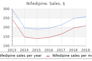
"Buy generic nifedipine 20 mg online, blood pressure ranges nhs."By: Danielle Marie Brander, MD - Assistant Professor of Medicine
- Member of the Duke Cancer Institute
 https://medicine.duke.edu/faculty/danielle-marie-brander-md
Buy cheap nifedipine on lineVascular dysfunction within the alphagalactosidase A-knockout mouse is an endothelial cell-, plasma membranebased defect. Alpha-galactosidase A deficiency accelerates atherosclerosis in mice with apolipoprotein E deficiency. Chapter 27 Renal Disease Caused by Inborn Errors of Metabolism, Storage Diseases, and Hemoglobinopathies 1271 121. Clinical, morphological, and molecular features of sialic acid storage illness manifesting in utero. Clinical, biochemical, and cytochemical research on a Japanese Salla illness case associated with a renal disorder. Newborn screening and early biochemical follow-up in combined methylmalonic aciduria and homocystinuria, cblC sort, and utility of methionine as a secondary screening analyte. Late-onset thrombocytic microangiopathy caused by cblC illness: Association with an element H mutation. Identification of the gene answerable for methylmalonic aciduria and homocystinuria, cblC type. Long-term end result in methylmalonic acidurias is influenced by the underlying defect (mut0, mut-, cblA, cblB). Long-term exposure of human proximal tubule cells to hydroxycobalamin[c-lactam] as a potential mannequin to research renal illness in methylmalonic acidurias. Combined liver-kidney transplant for the management of methylmalonic aciduria: A case report and evaluate of the literature. Renal sonographic findings of sort I glycogen storage illness in infancy and early childhood. The molecular foundation of glycogen storage disease sort 1a: Structure and performance evaluation of mutations in glucose-6-phosphatase. Glycogen storage illness kind I: Diagnosis, management, clinical course and outcome. Angiotensin mediates renal fibrosis in the nephropathy of glycogen storage disease sort Ia. Combined liver-kidney transplantation in glycogen storage illness Ia: A case past the guidelines. Neonatal gene remedy of glycogen storage disease sort Ia utilizing a feline immunodeficiency virus-based vector. Genetic classification and mutational spectrum of greater than 600 patients with a Zellweger syndrome spectrum disorder. Zellweger syndrome with unusual findings: Non-immune hydrops fetalis, dermal erythropoiesis and hypoplastic toe nails. Pyridoxamine lowers kidney crystals in experimental hyperoxaluria: A potential remedy for major hyperoxaluria. Long time period outcomes of liverkidney transplantation in kids with primary hyperoxaluria. Toward understanding renal Fanconi syndrome: Step by step advances by way of experimental models. Glycogen accumulation in the pars recta of the proximal tubule in Fanconi syndrome. A novel gene encoding an integral membrane protein is mutated in nephropathic cystinosis. Cysteamine therapy delays the progression of nephropathic cystinosis in late adolescents and adults. Nephropathic cystinosis in adults: Natural history and results of oral cysteamine therapy. Late-onset nephropathic cystinosis: Clinical presentation, outcome, and genotyping. Cystinosin, the protein defective in cystinosis, is a H(+)-driven lysosomal cystine transporter. Primary hyperoxaluria Type 1: Indications for screening and steering for diagnosis and remedy. Biochemical analyses are instrumental in figuring out the influence of mutations on holo and/or apo-forms and on the region(s) of alanine:glyoxylate aminotransferase variants associated with Primary Hyperoxaluria Type I. Molecular requirements for peroxisomal targeting of alanine-glyoxylate aminotransferase as an essential determinant in primary hyperoxaluria sort 1. The enzyme 4-hydroxy-2-oxoglutarate aldolase is poor in primary hyperoxaluria type 3. Chapter 27 Renal Disease Caused by Inborn Errors of Metabolism, Storage Diseases, and Hemoglobinopathies 1273 221. Inhibition of Na(+)-dependent transporters in cystine-loaded human renal cells: Electrophysiological studies on the Fanconi syndrome of cystinosis. Lysosomal cystine storage augments apoptosis in cultured human fibroblasts and renal tubular epithelial cells. Renal phenotype of the cystinosis mouse mannequin is dependent upon genetic background. Kidney preservation by bone marrow cell transplantation in hereditary nephropathy. Endosomal chloride-proton exchange rather than chloride conductance is crucial for renal endocytosis. Loss of chloride channel ClC-5 impairs endocytosis by defective trafficking of megalin and cubilin in kidney proximal tubules. Protein reabsorption in renal proximal tubulefunction and dysfunction in kidney pathophysiology. Effect of hydrochlorothiazide on urinary calcium excretion in dent disease: An uncontrolled trial. Tubulointerstitial nephritis related to a novel mitochondrial point mutation. Focal segmental glomerulosclerosis related to mitochondrial cytopathy: Report of two cases with particular emphasis on podocytes. Granular swollen epithelial cells: A histologic and diagnostic marker for mitochondrial nephropathy. Acquired Fanconi syndrome is an indolent dysfunction in the absence of overt a quantity of myeloma. Iminoglycinuria and hyperglycinuria are discrete human phenotypes ensuing from complex mutations in proline and glycine transporters. Molecular, biochemical, and genetic characterization of a female affected person with Lesch-Nyhan disease. Progressive morphologic renal modifications in the oculo-cerebro-renal syndrome of Lowe.
Purchase nifedipine 20mgAbout 80% of patients with excessive titers of antidonor cytotoxic antibodies in pretransplant crossmatch tests reject their kidney hyperacutely (520). One reason could also be low affinity, as some only react within the cold or dissociate after a number of washes. Hyperacute rejection does rarely occur within the absence of demonstrable antidonor antibody, presumably due to primed cytotoxic T cells current in the circulation on the time of transplantation. The first seen lesion within 30 minutes consists of lymphocytes attached to the arterial endothelium; after a couple of hours, the graft develops florid mononuclear infiltrate and necrosis. T-cell�mediated hyperacute rejection of mouse heart allografts has additionally been described within the absence of preexisting donor-reactive antibodies (522). Differential Diagnosis the differential diagnosis of hyperacute rejection includes perfusion injury and main vascular thrombosis. Perfusion harm has prominent lack of endothelium and rare thrombi however typically no important hemorrhage and necrosis. Clinical Course, Prognosis, Therapy, and Clinicopathologic Correlations Removal of the necrotic graft is often necessary to stop the development of systemic toxicity. In one case, a follow-up biopsy at 30 days showed decision of the glomerular thrombi (497). In another case, transplant glomerulopathy was evident at 39 days posttransplant (524). New drugs that block complement activation, similar to eculizumab, are underneath analysis (527). If the titer of antibodies diminishes to low or undetectable, transplantation has been safely undertaken, although antibodies had been beforehand current (525,530). In some sufferers, the antibodies return with both Chapter 29 Renal Transplant Pathology 1359 an episode of acute rejection or no instant impact on graft operate (accommodation). Detection of C4d in graft endothelium and the new solid-phase methods for detecting antidonor antibody have led to higher prognosis of this situation. Furthermore, deposition of immunoglobulin or C3 was not conspicuous in these cases. They advised that humoral rejection ought to be considered, despite unfavorable crossmatch earlier than transplantation and paucity of immunoglobulin deposition. These observations have been confirmed in lots of centers, and the criteria at the second are extensively accepted Banff consensus (82,461). Antibody induced graft injury, particularly acute, smoldering and chronic antibody-mediated rejection, can present concurrent T-cell mediated rejection (acute and/or chronic). In one collection, the imply day of onset was 15 � 11 days (earliest three days), not different from that of acute mobile rejection (14 � 10; earliest 6 days) (387). Late onset is commonly associated with iatrogenic or patient-initiated decreased immunosuppression (536,537). In one sequence, the majority fell inside the Banff "suspicious/borderline" range (Banff category 3) (388). However, neutrophils have been hardly ever (less than 3%) present in a collection from Vienna (388). Capillaritis may be present with little or no C4d deposition, which may observe a number of days later (549). Another research noted 53% of 17 patients had both fibrinoid necrosis (24%) or transmural arterial irritation (18%), or each (12%) (550). Tubules Evidence of acute tubular damage is widespread (loss of brush borders, thinning of cytoplasm, paucity of nuclei); in a single series, these have been present in 75% of instances (387). A: Transplant glomerulitis with mononuclear cells, neutrophils, and reactive endothelial cells. The capillary staining is crisp, linear, and steady, but also could have a finely granular pattern at high power, which extends into the lumen from the more linear deposits. Medullary vessels are typically optimistic and can be the one place of C4d positivity in some cases with marked edema and cortical injury (141,531). Intraluminal and interstitial C4d may also be seen, however is an artifact of fixation. In a series with serial biopsies of presensitized patients, C4d turned positive later after the capillaritis had been present for several days (549). This location suits with the identified capability of C4b to crosslink to nearby proteins at the website of complement activation. The covalent linkage of C4d to structural proteins might explain why C4d stays for several days after alloantibody disappears, since antibody binds to cell surface antigens that could be misplaced by modulation, shedding, or cell death. C4d can be detected on the floor of the endothelial cells and in intracytoplasmic vesicles by immunoelectron microscopy (554). Outcome at 1 year was not affected by C4d status at 7 days, regardless of the dearth of particular treatment. C4d deposition disappears a couple of days after treatment, supplied the antibody disappears, as judged by sporadic repeat biopsies and experimental research. C3d, produced by the classical pathway after C4b, has been instructed as an indicator of extra full complement activation. However, the usually excessive background of C3 deposition in the tubular basement membranes makes C3d much harder to interpret than C4d (558). These changes are extra evident in allografts that later develop transplant glomerulopathy (220,560). B: A glomerulus has one capillary plugged with fibrin (arrows) and one other filled with compacted red cells surrounded by a reactive endothelial cell (C); a 3rd loop has a quantity of platelets (arrowheads). Intact platelets are few, but microvesicles presumably derived from platelets are common (555). Endothelial cells present swelling, detachment, and enlargement of the subendothelial house with electron-lucent "fluffy" material and generally trapped purple cells (561). These changes are extra extreme and extensive than the endothelial swelling and apoptosis that happens in ischemic renal injury (560). After 2 to 4 weeks, the endothelial cells present cytoplasmic processes extending into the lumen and early multilayering of the basement membrane (560,561). These observations illustrate that electron microscopy can uncover signs of persistent rejection before they turn out to be more distinguished and detectable by commonplace light microscopy. In extreme instances, after 2 to 3 months, some capillaries are completely destroyed, with disappearance of the endothelial lining and remnants of the basement membrane; those who stay have a thickened, multilayered basement membrane (561). Histologic proof of acute tissue injury, including a number of of the next: - Microvascular irritation (g > 0c and/or ptc > 0) - Intimal or transmural arteritis (v > 0)d - Acute thrombotic microangiopathy, within the absence of another cause - Acute tubular harm, in the absence of any other obvious trigger 2. This has been included as an alternative criterion for antibody interplay with the vessels Table 29. Fortunately, other illnesses in the differential are unfavorable for C4d (141,461,479). Whether different donor antigen-expressing cells within the allograft may be focused (such as epithelial cells, clean muscle) is unknown. The nature of those antigens is unknown, excluding autoantibodies to the angiotensin 1 receptor (567).
Buy generic nifedipine 20 mg onlineThere is complete opacification of the left hemithorax, with a shift of the mediastinum to the left. Cavitation Cavitation is the presence of an area of radiolucency inside a mass lesion. Thymic tumours, thyroid masses and dermoid cysts are mostly situated in the anterior mediastinum, whereas neural lesions. Aneurysmal enlargement of the aorta or ventricle could produce masses within the center compartment of the mediastinum. It is useful in assessing lesions of the pleura and is particularly useful for localising loculated pleural effusions and guiding chest tube insertion (see Chapter 16). Fibrosis Localised fibrosis produces streaky shadows with proof of traction upon neighbouring buildings. Upper lobe fibrosis causes traction upon the trachea and elevation of the hilar vascular shadows. Generalised interstitial fibrosis produces a hazy shadowing with a fine reticular (netlike) or nodular sample (see Chapter 13). Diagram of lateral view of the chest, indicating the websites favoured by a few of the extra frequent mediastinal lots. Top: heart and major blood vessels, exhibiting the aorta curling over the bifurcation of the pulmonary trunk into left and proper pulmonary arteries (arrows). The aorta curls over the left main bronchus, which lies behind the left pulmonary artery. Pulmonary arteries are proven shaded, pulmonary veins are shown unshaded and bronchi are proven striped. The veins are utilized to the fronts of the arteries and bronchi and take a slightly different path to the respective lung segments. On the right, the order of buildings from entrance to again is vein�artery�bronchus; on the left, the pulmonary artery loops over the left upper lobe bronchus and descends behind, in order that the order is vein�bronchus�artery. Many giant vessels and an anterior sausage shape are seen; the trachea has not bifurcated (black circle). It is based on the concept that neoplastic cells have greater metabolic exercise and the next uptake of glucose than normal cells. It is also significantly helpful within the differential diagnosis of an indeterminate solitary pulmonary nodule. Calcification or lack of growth of the lesion over time means that the nodule is benign. If the patient is a smoker at high risk of most cancers however otherwise match, it could be advisable to proceed directly to surgical 56 Radiology of the chest resection of such a lesion, with out preoperative histological confirmation. False-negative findings can occur in tumours <1 cm and false-positive uptake can occur in inflammatory situations corresponding to tuberculosis, sarcoidosis, histoplasmosis and coccidioidomycosis. It ought to be studied in a systematic method and interpreted within the context of all scientific info. The more than likely prognosis is: A pneumonia B pneumonia with a parapneumonic effusion C mucus plugging of the left lower lobe bronchus D bronchial carcinoma E an inhaled overseas physique in the left lower lobe bronchus 4. The most likely trigger is a: A hiatus hernia B thymoma C oesophageal cyst D pericardial cyst E neurofibroma four. They are the commonest respiratory complaint, accounting for about 9% of all consultations normally follow. A child suffers about eight, and an grownup about four respiratory infections each year. No particular therapy is feasible for the frequent chilly, but signs are sometimes alleviated by use of paracetamol or aspirin. Pharyngitis Pharyngitis might happen as a half of the common chilly or as a separate sickness. A blood movie could show atypical mononuclear cells and the Monospot or heterophile antibody check is optimistic. Characteristically, sufferers with infectious mononucleosis develop a rash if given amoxicillin as remedy for pharyngitis. It could also be caused by about 200 completely different strains of viruses, including rhinoviruses, coronaviruses, respiratory syncytial, parainfluenza and influenza viruses. Infection is transmitted by droplet spread, and attack charges are highest in younger children attending faculty, who then transmit infection to their dad and mom and siblings at Respiratory Medicine Lecture Notes, Ninth Edition. Local extension of infection might lead to otitis media, tonsillitis or quinsy (peritonsillar abscess). Streptococcal infection may be sophisticated by glomerulonephritis or rheumatic fever, however these are rare these days. Antibiotic remedy of pharyngitis is normally solely given to extreme or complicated circumstances. Mycoplasma pneumoniae or Chlamydophila pneumoniae requires a tetracycline or macrolide antibiotic. A number of organisms may cause sinusitis, including respiratory viruses, Haemophilus influenzae, Streptococcus pneumoniae, Staphylococcus aureus and anaerobic bacteria. In continual sinusitis, X-rays may show mucosal thickening, opacification or the presence of a fluid degree in the sinus. Recurrent sinusitis may be accompanied by extra widespread respiratory tract an infection in patients with bronchiectasis attributable to cystic fibrosis, hypogammaglobulinaemia or ciliary dyskinesia. Post-nasal drip from sinusitis is irritating to the larynx and may cause a persistent cough. Croup Croup (acute laryngotracheobronchitis) is often attributable to viruses similar to parainfluenza virus, respiratory syncytial virus, influenza A and B, rhinoviruses, adenovirus and measles. Characteristically, a child develops a harsh barking cough with an upper respiratory infection, which can progress to stridor. Often, no remedy is required, however some children develop more extreme lower respiratory infections and progressive respiratory misery, requiring intubation and ventilation. Oral prednisolone is typically beneficial in extreme croup and nebulised high-dose budesonide may be related to more speedy restoration in much less severely affected sufferers. Pertussis Pertussis (whooping cough) is an infectious disease of the respiratory tract caused by Bordetella pertussis. Influenza virus type A undergoes frequent spontaneous modifications in its haemagglutinin and neuraminidase floor antigens. Influenza is extremely infectious, so that every one members of a household typically turn into sick collectively. It is usually a self-limiting sickness, however it can be difficult by bronchitis, otitis media and secondary bacterial pneumonia. The analysis of influenza can be confirmed by immunofluorescent microscopy of nasal secretions or by serology. Oseltamivir and zanamivir are drugs that reduce the replication of influenza viruses by inhibiting viral neuraminidase. These medicine need to be given inside forty eight hours of the onset of symptoms to be efficient. They reduce the duration of illness by about 1 day and they could cut back problems in at-risk patients with extreme influenza.
Cheap nifedipine lineThe endothelial cell in ischemic acute kidney damage: Implications for acute and continual perform. Endothelial activation and circulating markers of endothelial activation in kidney disease. Preservation of peritubular capillary endothelial integrity and growing pericytes may be important to restoration from postischemic acute kidney damage. Guanosine supplementation reduces apoptosis and protects renal perform within the setting of ischemic damage. Chemical inhibitor of nonapoptotic cell dying with therapeutic potential for ischemic brain harm. Rip1 (Receptorinteracting protein kinase 1) mediates necroptosis and contributes to renal ischemia/reperfusion injury. Protective impact of T cell depletion in murine renal ischemia-reperfusion harm. Effects of mixed T- and B-cell deficiency on murine ischemia reperfusion damage. Compartmentalization of neutrophils in the kidney and lung following acute ischemic kidney harm. Chapter 26 Ischemic and Toxic Acute Tubular Injury and Other Ischemic Renal Injuries 1217 400. Calmodulin regulates fodrin susceptibility to cleavage by calcium-dependent protease I. Redistribution and dysfunction of integrins in cultured renal epithelial cells uncovered to oxidative stress. Functional and cytoskeletal modifications induced by sublethal damage in proximal tubular epithelial cells. Effects of integrins on proliferation and apoptosis of renal epithelial cells after acute injury. Heat shock protein expression is highly delicate to ischemia-reperfusion damage in rat kidneys. Characterization of denatured protein inducers of the heat shock (stress) response in Xenopus laevis oocytes. Ischemic acute renal failure induces differential expression of small warmth shock proteins. Heat-shock protein 25 induction and redistribution during actin reorganization after renal ischemia. Hsp72 expression enhances survival in adenosine triphosphate-depleted renal epithelial cells. Preconditioning with sodium arsenite inhibits apoptotic cell dying in rat kidney with ischemia/reperfusion or cyclosporine-induced Injuries. Altered cholesterol localization and caveolin expression through the evolution of acute renal failure. In vivo and in vitro fashions show a job for caveolin-1 in the pathogenesis of ischaemic acute renal failure. Lipoxins: Potential antiinflammatory, proresolution, and antifibrotic mediators in renal disease. Shedding of kidney harm molecule-1, a putative adhesion protein concerned in renal regeneration. Epidermal development factor accelerates useful recovery from ischaemic acute tubular necrosis in the rat: Role of the epidermal progress issue receptor. Hepatocyte development factor accelerates restoration from acute ischemic renal harm in rats. Blockage of tubular epithelial to myofibroblast transition by hepatocyte development issue prevents renal interstitial fibrosis. Restoration of tubular epithelial cells during restore of the postischemic kidney occurs independently of bone marrow-derived stem cells. Mesenchymal stem cells contribute to the renal repair of acute tubular epithelial harm. Mesenchymal stem cells are renotropic, helping to repair the kidney and improve function in acute renal failure. Kidney graft function determines endothelial progenitor cell number in renal transplant recipients. Hematopoietic stem cells contribute to the regeneration of renal tubules after renal ischemia-reperfusion damage in mice. Hematopoietic stem cell mobilization remedy accelerates restoration of renal function independent of stem cell contribution. Bone marrow stem cells contribute to restore of the ischemically injured renal tubule. Administered mesenchymal stem cells enhance restoration from ischemia/reperfusion-induced acute renal failure in rats. Administered mesenchymal stem cells defend against ischemic acute renal failure via differentiationindependent mechanisms. Aminoglycoside antibiotics bind to protein disulfide isomerase and inhibit its chaperone exercise. Gentamicin binds to the lectin website of calreticulin and inhibits its chaperone exercise. Antioxidant S-allylcysteine prevents gentamicin-induced oxidative stress and renal injury. Involvement of reactive oxygen species on gentamicin-induced mesangial cell activation. Selective membrane toxicity of the polyene antibiotics: Studies on natural membranes. Amphotericin B nephrotoxicity: the antagonistic penalties of altered membrane properties. Direct vasoconstriction as a possible trigger for amphotericin B-induced nephrotoxicity in rats. The impact of thromboxane A2 receptor antagonism on amphotericin B-induced renal vasoconstriction within the rat. Mechanisms of amphotericin B-induced discount of the glomerular filtration price: A micropuncture examine. Prediction of nephrotoxicant motion and identification of candidate toxicity-related biomarkers. Quantitative gene expression analysis in a nonhuman primate model of antibiotic-induced nephrotoxicity. Drug-induced renal failure: update on new medicines and distinctive mechanisms of nephrotoxicity. Cytotoxicity of antiviral nucleotides adefovir and cidofovir is induced by the expression of human renal organic anion transporter 1. Multidrug-resistance protein 5 is a multispecific organic anion transporter able to transport nucleotide analogs. The antiviral nucleotide analogs cidofovir and adefovir are novel substrates for human and rat renal natural anion transporter 1. Pharmacokinetics of lamivudine administered alone and with trimethoprim-sulfamethoxazole.
Discount nifedipine 20mg without prescriptionTubulitis, tubular cell damage, regenerative epithelial adjustments, and variable numbers of casts are seen. By immunofluorescence, deposits of immune complexes are absent from glomeruli or tubules. Tubulointerstitial nephropathy associated with monoclonal gammopathies is reviewed in Chapter 22. The mechanism by which inflammatory cells induce fibrosis has been reviewed (45,forty six,438) and might be mentioned right here briefly. Interstitial lymphocytes interact with monocytes/macrophages, with endothelial cells, and presumably with tubular epithelial cells (448) in antigen presentation, resulting in a delayed-type cellmediated reaction. Whether a diffuse cellular infiltrate or a granulomatous inflammation develops can be determined by the interaction of cytokines brought into the interstitial microenvironment by T cells (446). The mechanism of interstitial inflammation includes a number of biologic events, as discussed previously. Activated T cells, monocytes/macrophages, and renal tubular epithelial cells release chemokines and cytokines. Therefore, activated/injured tubular epithelial cells in interstitial nephritis, in turn, may further appeal to macrophages into the kidney and interstitium, aggravating the disease process (35,36). The nephropathies caused by persistent exposure to probably the most plentiful toxic metals-lead, cadmium, and mercury-are thought-about here in some detail; different nephropathies associated with heavy metal publicity are mentioned briefly. Diagnosing chronic heavy metallic exposure� related nephropathy is kind of difficult, and the condition is probably frequently missed. Contaminating food provide (such as seafood) and probably also drinking water (possible well water contamination in areas of fracking [hydraulic fracturing during pure gas/petroleum exploration]) is a rising concern and can doubtlessly be the supply of low-level chronic heavy metallic publicity to certain populations (455). Lead Nephropathy Lead publicity within the form of inhaled fumes and dirt is an occupational illness for industrial staff. Soil and paints containing lead are sources of lead publicity, notably for children (457). Absorbed lead is widely distributed, however the principal sites of long-term storage are the bones, by which 94% of the lead within the body is found. This storage web site constitutes a slow-exchange pool, and the biologic half-life of lead in bone is about sixteen years (458). Another 4% is present in the blood, tissue fluids, and gentle tissues, and these represent a rapid-exchange pool. The remaining 2% is distributed between actively exchanging elements of the skeleton and soft tissues. An epidemic of childhood lead poisoning in Queensland, Australia, established lead nephropathy as a acknowledged medical and pathologic entity (460,461). The check suggests lead nephropathy if excretion of lead is larger than 650 mg in 24 hours. Because the half-life of circulating lead is about 1 month, the test displays only recent exposure (457), and the result could be normal for sufferers with continual lead toxicity (457,462). The lead focus may also be measured in tissues (primarily bone) by x-ray fluorescence and neutron activation analyses (458). Many research have confirmed a relationship between lead exposure and persistent renal disease (462�464). On the other hand, a study from Taiwan lately examined the impact of environmental lead publicity on the progression of continual renal disease and located that even low-level environmental lead exposure is related to progressive renal insufficiency (463). One hundred and twenty-one patients had been included in the study with a baseline creatinine degree between 1. Seventeen patients doubled their baseline Scr quantity within the follow-up period of forty eight months. Blood lead ranges and physique lead burden at baseline have been the most important risk elements to predict development of renal insufficiency. None of the sufferers had a history of lead exposure, and all of them had blood lead ranges and body lead burden above acceptable ranges (463). Because of the nature of their study, they may not conclude whether lead exposure resulted in impaired renal operate or whether or not impaired renal perform caused elevated focus of lead in the blood. Chronic lead intoxication is manifested by proximal tubular defects, and decreased glucose reabsorptive capacity is an early indicator of tubular cell damage (467). Most Clinical Presentation patients have recurrent gout, hyperuricemia, and hypertension (468). Whether hypertension and hyperuricemia are caused by lead publicity, however, is controversial (457,459). However, several studies help the reality that lead could cause decreased renal uric acid excretion and uric acid deposition in the kidney, which can be one essential issue within the improvement of persistent lead nephropathy (468). Also, long-term accumulation of lead within the body is probably an unbiased risk issue for the development of hypertension (468). Pathologic Findings the kidneys are shrunk, show a finely granular surface with discount of the cortex, and will weigh one third of regular (461). There is variable multifocal tubular atrophy, tubular loss, and interstitial fibrosis (461,469). Glomeruli are regular (469), and arteries and arterioles show medial thickening and luminal narrowing, in all probability associated to hypertension. Lead in fluids is certain to lead-binding proteins and is taken up by epithelial cells by membrane binding and probably by passive transport; absorbed lead accumulates preferentially in proximal tubular cells (469,470). A cleavage product of 2-microglobulin is the principal component of complexed lead that makes Pb2+ out there to enzymes (-aminolevulinic acid dehydrase) and mediates intranuclear transport and chromatin binding, leading to adjustments in gene expression. Lead interacts with renal membranes and enzymes; disrupts power manufacturing, calcium metabolism, and glucose homeostasis; and interferes with ion transport. Oxidative stress more than likely plays a major role in the pathogenesis as a end result of serum ranges of oxidative stress markers show a close correlation with lead exposure ranges (470). It seems that urine degree of alphaglutathione S-transferase, a marker of proximal tubular harm, could also be an early marker of lead nephrotoxicity (470). Cadmium Nephropathy Cadmium publicity from inhalation of cadmium oxide dust or cadmium fumes is an occupational sickness (456) that happens in the manufacture of pigments, plastics, electric storage batteries, and metal alloys. In the general inhabitants, exposure happens by the oral route by way of contaminated water or meals or inhalation of indoor dust contaminated with cadmium (471). Cigarette smoking is one other potential source of publicity, as a end result of cadmium aerosol, produced throughout smoking, facilitates absorption of the steel. Cadmium toxicity is manifested by elevated excretion of high and low molecular weight proteins, corresponding to 2-microglobulin (475), kidney-derived antigens, enzymes, prostanoids, glycosaminoglycans, sialic acid, glucose, and amino acids or the total complement of substances seen in the Fanconi syndrome (476). Subclinical modifications in tubular operate additionally occur in the common inhabitants above a threshold excretion of urinary cadmium of 2 mg in 24 hours (476). Once manifested, renal harm tends to be progressive, even if exposure is discontinued (477,478). In addition to irreversible dysfunction of proximal tubules, excess cadmium publicity can also be known to cause hypercalciuria, nephrolithiasis, and osteomalacia. It has been demonstrated that for each doubling of urinary cadmium focus, the relative danger for mortality will increase by 17% (473). Nogawa (479) reported low-level extended environmental publicity to cadmium via contaminated water within the Kakehashi River basin in Japan.
Black Elder (Elderberry). Nifedipine. - Are there safety concerns?
- What is Elderberry?
- Are there any interactions with medications?
- Cancer, constipation, nerve pain, chronic fatigue syndrome (CFS), hayfever, HIV/AIDS, and other conditions.
- How does Elderberry work?
Source: http://www.rxlist.com/script/main/art.asp?articlekey=96444
Purchase 20mg nifedipine free shippingWith timely renal alternative therapy, renal perform may recover sufficiently to permit patients to turn out to be dialysis independent after a interval of 1 to three months, and renal perform may proceed to enhance over a period of 1 to 2 years. No specific therapeutic approaches have been profitable, although anticoagulants, beta-blockers, cytotoxic medicine, and mannitol have all been tried. B: Gross specimen of a kidney with extensive areas of pallor representing the necrotic cortex with intervening areas of congested nonnecrotic parenchyma. Light Microscopy By gentle microscopy, the findings are very similar to those seen with ischemic infarcts. Patients biopsied late within the course have bland parenchymal fibrosis/sclerosis (689). The majority of infarcts in adults are often clinically silent and are incessantly brought on by sudden and complete arterial blockage by emboli, doubtlessly originating from ventricular thrombi or from vegetations on coronary heart valves with verrucous or infective endocarditis or because of atheroemboli (see below). Less generally, arterial obstruction is produced by thrombosis owing to adjustments within the wall of the vessel associated with atherosclerosis, systemic sclerosis, malignant hypertension, polyarteritis, or aneurysm formation because of dysplastic illness of the renal artery. Renal infarction has also not often been described on account of cocaine use (690,691). Cases have also been described with fibromuscular dysplasia involving the renal arteries. Loin or renal fossa pain, hematuria, and/or pyrexia may counsel an infection or renal calculi. Renal infarction incessantly goes undetected, however, significantly if the area of infarction is small. Large infarcts may be associated with loin pain followed by hematuria and transient proteinuria. Arterial occlusion is extra regularly seen as a reason for renal infarction than venous occlusion. It may result from embolism, thrombosis, or vessel wall injury which accompanies malignant hypertension or systemic vasculitis. The gross appearance of a renal infarct is determined by the age of the lesion, the size of the vessel obstructed, and the presence or absence of infection. Initially, the infarct is purple and pyramidal in form, with the apex toward the obstructed artery. As necrotic tissue is removed and replaced by collagenous tissue, the area of infarct shrinks and ultimately becomes a V-shaped scar. The medulla is generally spared in renal infarction, and the lesions are confined to the cortex. Infarcts resulting from septic embolization are associated with the presence of liquefactive necrosis and abscess formation as a end result of digestion by leukocytic enzymes. Light Microscopy Histologically, sterile infarcts present the findings of classic coagulative necrosis. As the lesion progresses, the central necrotic space becomes smaller, and group and regeneration occur around the periphery, with eventual collapse of the central necrotic space and replacement by collagenous scarring. As talked about beforehand, the lesions are typically confined to the cortical tissue, and the medulla is spared. This image helps distinguish scars ensuing from infarction from those brought on by reflux or pyelonephritis, by which medullary involvement is prominent. Whether infarction occurs within the kidney because of venous thrombosis is dependent upon the completeness of the occlusion and the speed at which thrombosis and occlusion take place. The emboli are normally derived from Venous Infarction Venous obstruction as a reason for infarction is way less widespread than arterial obstruction. It is seen in infancy as a complication of severe dehydration stemming from diarrheal diseases. Note utterly necrotic tubules (arrows) and relatively intact tubules with individual cell necrosis (arrowheads) containing necrotic debris. Transient azotaemia is associated with a high risk of dying in hospitalized sufferers. Acute Kidney Injury Network: Report of an initiative to enhance outcomes in acute kidney injury. Lower nephron nephrosis: the renal lesions of the crush syndrome, of burns, transfusions and other conditions affecting the lower segments of the nephrons. The pathogenesis of acute renal failure associated with traumatic and poisonous damage: Renal ischemia, nephrotoxic injury and the ischemic episode. Acute Anuria: A Study Based on Renal Function Tests and Aspiration Biopsy of the Kidney. Renal interstitial pressure in regular and in anuric man: Based on wedged renal vein pressure. Acute renal failure due to nephrotoxins: Renal hemodynamic and angiographic studies in man. Acute oliguric renal failure in man: Evidence for preferential renal cortical ischemia. When obstruction is full, distal areas of infarction and necrosis are evident. In early-stage lesions, the crystals are surrounded by fibrin, occasionally with related eosinophils. In older embolic lesions, organization is evident, and the crystals are surrounded by fibrous tissue. Renal atheroemboli are frequently a part of more generalized atheroembolic disease, ensuing from multiple showers of cholesterolcontaining microemboli in many organs, including the retina, brain, pancreas, and, in particular, the muscles and skin of the legs in addition to the kidney. Multisystem involvement often mimics systemic vasculitides, similar to microscopic polyarteritis. The presence of eosinophilia, hypocomplementemia, and generally eosinophiluria further complicates the scientific recognition of this disease. Tenofovir-associated acute and chronic kidney disease: A case of a quantity of drug interactions. Tenofovir nephrotoxicity: Acute tubular necrosis with distinctive scientific, pathological, and mitochondrial abnormalities. Risk factors for nephrotoxicity in aged patients receiving once-daily aminoglycosides. Aminoglycoside-associated extreme renal failure in patients with a number of myeloma handled with thalidomide. Deafness and acute tubular necrosis following parenteral administration of neomycin. Toxicity of amphotericin B plus flucytosine in 194 sufferers with cryptococcal meningitis. The epidemiology of nephrotoxicity related to standard amphotericin B remedy. Effect of amphotericin-B on water and urea transport within the inside medullary collecting duct. Drug-induced nephrotoxicity caused by amphotericin B lipid complex and liposomal amphotericin B: A review and meta-analysis. Nephrotoxicity of beta-lactam antibiotics: mechanisms and techniques for prevention.
Syndromes - Chest x-ray
- Infections (such as strep throat, hepatitis, or mononucleosis)
- FSH
- Loss of appetite
- Confusion
- Blood in the urine
- Body lice
- Retinal detachment
- Thoracentesis
- Creatinine clearance - urine test
Order nifedipine cheapThe destiny of glomerular mesangial IgA deposition within the donated kidney after allograft transplantation. Transplantation and 2-year follow-up of kidneys procured from a cadaver donor with a historical past of lupus nephritis. Successful transplantation of a cadaveric kidney with post-infectious glomerulonephritis. Transmission and determination of type I membranoproliferative glomerulonephritis in recipients of cadaveric renal allografts. Genome-wide gene-expression patterns of donor kidney biopsies distinguish primary allograft perform. Early loss of renal transcripts in kidney allografts: relationship to the development of histologic lesions and alloimmune effector mechanisms. Gene-expression profiles and age of donor kidney biopsies obtained earlier than transplantation distinguish medium term graft function. Laparoscopic donor nephrectomy gene expression profiling reveals upregulation of stress and ischemia associated genes in comparison with management kidneys. Comparing molecular assessment of implantation biopsies with histologic and demographic risk assessment. Cohort research of the prognostic significance of acute transplant glomerulitis in acutely rejecting renal allografts. The mobile lesion of humoral rejection: predominant recruitment of monocytes to peritubular and glomerular capillaries. Infiltrating cell sorts in transplant glomerulitis: relationship to peritubular capillary C4d deposition. Pathologic features of acute renal allograft rejection related to donor-specific antibody. Immunopathology of renal allograft rejection analyzed with monoclonal antibodies to mononuclear cell markers. A critical appraisal of strategies to grade transplant glomerulitis in renal allograft biopsies. Glomerular irritation in renal allografts biopsies after the primary yr: cell types and relationship with antibody-mediated rejection and graft consequence. Segmental localization and quantitative characteristics of tubulitis in kidney biopsies from sufferers undergoing acute rejection. A new diagnostic algorithm for antibody-mediated microcirculation inflammation in kidney transplants. Immunohistological evaluation of sequential renal biopsies from patients with acute renal rejection. Results from a human renal allograft tolerance trial evaluating T-cell depletion with alemtuzumab mixed with deoxyspergualin. Plasma cell densities and glomerular filtration rates predict renal allograft outcomes following acute rejection. Concurrent acute mobile rejection is an independent threat issue for renal allograft failure in sufferers with C4d-positive antibody-mediated rejection. The classification and remedy of antibodymediated renal allograft damage: where can we stand Factors contributing to acute rejection in renal transplantation: the position of noncompliance. Organ procurement and transplantation community and scientific registry of transplant recipients 2010 annual information report. Detection of the complement degradation product C4d in renal allografts: diagnostic and therapeutic implications. Renal allograft biopsies in the era of C4d staining: the need for change in the Banff classification system. Antibody-mediated vascular rejection of kidney allografts: a population-based study. Pathological modifications in 37 human renal homotransplants treated with immunosuppressive medication. A case of persistent acute allograft glomerulopathy with long-standing secure renal operate. Acute renal allograft rejections with major interstitial oedema and plasma cell-rich infiltrates: high gamma-interferon expression and poor scientific outcome. Acute rejection and graft survival in renal transplanted patients with viral illnesses. Acute interstitial nephritis of plasma cells: a brand new cause for renal allograft loss. The medical and pathologic implications of plasmacytic infiltrates in percutaneous renal allograft biopsies. Immunohistochemical analysis of the interstitial mast cells in acute rejection of human renal allografts. The prognostic significance of particular arterial lesions in acute renal allograft rejection. Abundance of interstitial eosinophils in renal allografts is associated with vascular rejection. Significance of interstitial lesions as the early indicator for acute vascular rejection in human renal allografts. Morphology of cyclosporine nephrotoxicity and acute rejection in patients immunosuppressed with cyclosporine and prednisone. Interstitial rejection, vascular rejection, and diffuse thrombosis of renal allografts. An evaluation of the Banff classification of early renal allograft biopsies and correlation with end result. Peritubular capillaritis in early renal allograft is related to the development of continual rejection and continual allograft nephropathy. Expression and tissue localization of donor-specific complement C3 synthesized in human renal allografts. Interleukin 2 mediates stimulation of complement C3 biosynthesis in human proximal tubular epithelial cells. Effect of human recombinant cytokines on the induction of macrophage procoagulant exercise. Humoral rejection in kidney transplantation: new concepts in prognosis and remedy. Transplant glomerulopathy: ultrastructural abnormalities occur early in longitudinal analysis of protocol biopsies. Postcapillary venule-like transformation of peritubular capillaries in acute renal allograft rejection. Diagnostic significance of peritubular capillary basement membrane multilaminations in kidney allografts: old ideas revisited. Quantitative detection of immune activation transcripts as a diagnostic tool in kidney transplantation.
Purchase 30 mg nifedipine with mastercardInterleukin-17 activates human renal epithelial cells in vitro and is expressed throughout renal allograft rejection. Renal allograft rejection: betachemokine involvement within the development of tubulitis. Renal transplantation: examination of the regulation of chemokine binding throughout acute rejection. Two-color move cytometry and practical evaluation of lymphocytes cultured from human renal allografts: identification of a Leu-2+3+ subpopulation. Topographical distribution of inflammatory leukocyte subsets in acute cellular rejection of a kidney allograft. Characterization of T cells expressing the / antigen receptor in human renal allografts. Dominant rearrangements of TcR beta genes and persistence of dominant rearrangements in serial biopsies. Clonality evaluation of T cells mediating acute and continual rejection in kidney allografts. T-cell receptor variable gene evaluation of renal allograft-infiltrating cells in biopsy specimens using a nonradioisotopic micromethod. Matching T-cell receptors identified in renal biopsies and urine at the time of acute rejection in pediatric renal transplant sufferers. Infiltration of perforin-positive mononuclear cells into the rejected kidney allograft. Increased accuracy of renal allograft rejection analysis utilizing mixed perforin, granzyme B, and Fas ligand fine-needle aspiration immunocytology. Induction of Fas-mediated apoptosis in a human renal epithelial cell line by interferon-gamma: involvement of Fas-mediated apoptosis in acute renal rejection. Expression of granzyme A and B proteins by cytotoxic lymphocytes concerned in acute renal allograft rejection. Functional analysis of lymphocyte subsets and clones infiltrating renal allografts. The regulatory/cytotoxic graft-infiltrating T cells differentiate renal allograft borderline change from acute rejection. Processing and presentation of self- and non-self foreign antigens by the renal proximal tubule. Tumor necrosis factor alpha in human kidney transplant rejection-analysis by in situ hybridization. Tubular expression of intercellular adhesion molecule-1 throughout renal allograft rejection. Variation in expression of endothelial adhesion molecules in pretransplant and transplanted kidneys- correlation with intragraft events. Alloantibody- and T cell-mediated immunity in the pathogenesis of transplant arteriosclerosis: lack of development to sclerotic lesions in B cell-deficient mice. Ultrastructural evidence of cell-mediated endothelial cell injury in cardiac transplantrelated accelerated arteriosclerosis. Presence of FoxP3+ regulatory T Cells predicts end result of subclinical rejection of renal allografts. Intragraft regulatory T cells in protocol biopsies retain foxp3 demethylation and are protecting biomarkers for kidney graft end result. In situ expression of tumor necrosis factor-alpha, interferon-gamma, and interleukin-2 receptors in renal allograft biopsies. Expression of inducible lymphocyte costimulatory molecules in human renal allograft. Cytokine and cytotoxic molecule gene expression decided in peripheral blood mononuclear cells in the analysis of acute renal rejection. Diagnostic and predictive value of an immunohistochemical profile in asymptomatic acute rejection of renal allografts. Immunohistological studies with A1-3, a monoclonal antibody to activated human monocytes and macrophages. Overexpression of soluble fas attenuates transplant arteriosclerosis in rat aortic allografts. Vascular endothelial growth issue expression and cyclosporine toxicity in renal allograft rejection. Expression of urokinase plasminogen activator and its receptor throughout acute renal allograft rejection. A morphological and immunohistological research on "zero-hour" and follow-up biopsies with special emphasis on mobile infiltrates and adhesion molecules. Renal allograft rejection- in situ demonstration of cytotoxic intratubular cells. Post-transplant renal tubulitis: the recruitment, differentiation and persistence of intra-epithelial T cells. De novo induction of endothelial L-selectin ligands during kidney allograft rejection. Identification of platelet-derived growth issue A and B chains in human renal vascular rejection. Pattern of endothelin immunostaining throughout rejection episodes after kidney transplantation. Typing of intraglomerular mononuclear cells associated with transplant glomerular rejection. Renal allograft involvement by Epstein-Barr virus related post-transplant lymphoproliferative illness. Clinical significance of renal biopsies exhibiting concurrent acute rejection and tacrolimus-associated tubular vacuolization.
[newline]Endothelial C4d deposition is related to inferior kidney allograft outcome independently of cellular rejection. Monocytes and peritubular capillary C4d deposition in acute renal allograft rejection. Comparison between bortezomib and rituximab in the therapy of antibody-mediated renal allograft rejection. Banff criteria as predictors of consequence following acute renal allograft rejection. Correlation between Banff classification, acute renal rejection scores and reversal of rejection. Is there any correlation between pathologic modifications for acute rejection in kidney transplantation (Banff 97) and graft perform Acute renal allograft rejection with intimal arteritis: histologic predictors of response to remedy and graft survival. Percutaneous needle biopsies of renal allografts: the connection between morphological changes current in the biopsies and subsequent allograft function. The predictive value of percutaneous biopsies from human renal allografts with early impaired operate. Clinical implications of the diagnosis of renal allograft infarction by percutaneous biopsy. Donor-transmitted adenovirus an infection causing kidney allograft nephritis and graft loss. Reflections on use of the renal biopsy because the "gold commonplace" in distinguishing transplant rejection from cyclosporine nephrotoxicity.
Generic nifedipine 30 mg otcThe endothelium is reactive (expanded endoplasmic reticulum) with loss of fenestrations. A subsequent comprehensive study by Liapis in contrast native and transplant kidneys (224). The specificity of those ultrastructural options was documented in indication biopsies taken within 3 months of transplantation (644). B Chapter 29 Renal Transplant Pathology 1379 glomerulopathy was sturdy (60% of C4d+ and 50% of cases with glomerulopathy). Scanning electron microscopy reveals endothelial cell injury, disorganization of the endothelium, and gaps between endothelial cells, typically with leukocytes and platelets. The thickened arterial intima consists of clean muscle cells, collagen fibrils, basement membrane materials, and a loose amorphous electron-lucent ground substance (642). Scattered lymphocytes and macrophages are present, the latter generally crammed with fantastic lipid droplets, similar to the froth cells by mild microscopy. With time, the cellularity diminishes and the quantity of collagen will increase (164,648). Biopsies with focal/diffuse C4d positivity showed increased expression of genes associated to the immune response, interferon- and rejection-induced, cytotoxic T-cell, macrophage-associated, and endothelial cell transcripts. Microarrays confirmed few differentially expressed genes between paired biopsies from recipients before and after the prognosis of transplant glomerulopathy. However, the glomerulopathy group had considerably altered expression for higher than 2000 genes at 4 to 24 months posttransplantation compared with the controls (no glomerulopathy or negative crossmatch). Molecular Studies Etiology and Pathogenesis Endothelium is the main, but perhaps not the only, goal of persistent rejection mediated by antibodies. Presumably, this is related to differences in the resistance of the endothelium (accommodation) or probably difference in practical activity of antibodies themselves. Whatever the mechanism, the endothelium responds by repeated synthesis of basement membrane, analogous to the response of other basement membrane producing cells. Risk Factors, Prognosis, and Differential Diagnosis Transplant Glomerulopathy the median time of prognosis of transplant glomerulopathy by indication biopsies is 5 to eight years (422,634). Transplant glomerulopathy is usually preceded by transplant glomerulitis, and glomerulitis coincides with glomerulopathy in roughly 50% of instances. Transplant glomerulopathy has a poor prognosis, significantly when accompanied by C4d deposition (419,655,656). In biopsies 10 years or more after transplantation, a majority (70%) of grafts with transplant glomerulopathy and C4d deposition failed within 1 12 months of biopsy, a a lot worse failure price than grafts with transplant glomerulopathy without C4d (15%) or grafts with C4d and no glomerulopathy (less than 5%). Among these in whom subclinical transplant glomerulopathy was detected in a 1-year protocol biopsy, the 3-year graft survival was 85%, worse than these with normal biopsies or only interstitial fibrosis (657,658). Proteinuria itself is associated with decreased graft survival in proportion to the diploma of proteinuria (660,661). The rate of graft loss in the presence of 1 g/d of proteinuria is 25% in the first 12 months and 62% by 5 years (662). Proteinuria because of recurrent or de novo glomerulonephritis had a greater prognosis (17% graft loss in 5 years). In any case, it is necessary to distinguish these mechanisms, as they might have different prognostic and therapeutic implications. The different main diagnoses to consider are recurrent or de novo glomerular illness. If immune advanced deposits are more than occasional or if in a subepithelial location, recurrent or de novo glomerulonephritis ought to be suspected. More severe arteriosclerosis was considerably related to capillaritis, glomerulitis, and interstitial irritation (628). Transplant arteriopathy is rare within the first 6 months after transplantation, but will increase to 36% to 41% in protocol biopsies at 1 to 2 years (658,668). Rarely, persistent arteriopathy has been reported as early as 1 month posttransplant (669). In nonhuman primates on little or no immunosuppression, the lesions can develop within 2 to three months (164,670). In the previous, arteriopathy influenced long-term survival greater than interstitial fibrosis (671). However, a current giant series showed no prognostic effect of arteriopathy in 1-year biopsies, separate from interstitial fibrosis, inflammation, or glomerulopathy, a discovering attributed to reduced severity of arteriopathy under current drug regimens (658). Gibson was a strong advocate for this otherwise neglected lesion and proposed a scoring system, which has since been included into the Banff system (622). Capillaritis was strongly associated with glomerulitis and C4d+: 24% of C4d adverse biopsies in contrast with 75% of C4d+ biopsies showed capillaritis. Nonetheless, diffuse capillaritis in early protocol biopsies had important adverse prognostic impact when it comes to glomerular filtration fee 2 years later. Capillaritis without C4d has been demonstrated in 3-month protocol biopsies in presensitized sufferers and is a threat issue for transplant glomerulopathy at 1 year (492,673). C4d deposition was a good stronger risk issue for later transplant glomerulopathy (up to 18-fold), but capillaritis remained predictive even after correction for C4d standing (up to sixfold). Another examine principally in presensitized patients showed indication biopsies with capillaritis in biopsies taken earlier than three months had a greater price of graft loss, particularly when not treated (644). Several elements contribute to the heterogeneity: the edge of "C4d-negativity" used (less than 50%, lower than 10% or 0%), the approach (fixed paraffin sections vs. There are vital therapeutic implications if C4d negativity implies complement-independent mechanisms being answerable for the harm. Alternatively, C4d deposition could by some means protect the endothelium or be irrelevant to the injury. The criteria for C4d unfavorable ("complement poor") have been proposed in the 2013 Banff consensus report (481). Because of the increased use of protocol biopsies, a fifth category has been identified (although not yet integrated into Banff). The Banff classification terms this state "C4d deposition with out evidence of energetic rejection" and recommends careful monitoring of sufferers with this sample (155). Regele showed that patients with C4d+ biopsies in the first year posttransplant had a better frequency of transplant glomerulopathy in later biopsies (554). A related sequence was famous by Loupy in 3-month protocol biopsies in sensitized patients, during which C4d+ or capillaritis predicted transplant glomerulopathy 9 months later. Further research are wanted to identify the danger factors for progression through the stages and the optimal therapy. The course of begins with antibody manufacturing, followed by C4d fixation in the tissue (if enough quantities of complement-fixing antibodies are formed). The inevitability of development has not been proved in people, although these stages happen in nonhuman primates with out immunosuppression. Patients with progressive loss of renal function commonly have hypertension and proteinuria, typically within the nephrotic range (640).
References - Harle LK, Maggio M, Shahani S, et al: Endocrine complications of androgendeprivation therapy in men with prostate cancer, Clin Adv Hematol Oncol 4(9):687n696, 2006.
- Shaikh N, Ewing AL, Bhatnagar S, et al: Risk of renal scarring in children with a first urinary tract infection: a systematic review, Pediatrics 126:1084- 1091, 2010.
- Bukowski TP, Chakrabarty A, Powell IJ, et al: Acquired rectourethral fistula: methods of repair, J Urol 153(3 Pt 1):730n733, 1995.
- Pansadoro V: Surgery illustrated: surgical atlas: the posterior lumbotomy, BJU Int 95(7):1121-1131, 2005.
- Kamat AM, Dinney CPN, Gee JR, et al: Micropapillary bladder cancer, Cancer 110:62n67, 2007.
|
|


