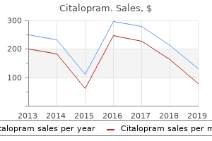
"Buy citalopram 40mg with mastercard, treatment 4s syndrome."By: Danielle Marie Brander, MD - Assistant Professor of Medicine
- Member of the Duke Cancer Institute
 https://medicine.duke.edu/faculty/danielle-marie-brander-md
Best 40mg citalopramThe technic for insertion of femoral head prosthesis by the straight anterior or Hueter method. Treatment of femoro-acetabular impingement with surgical dislocation and debridement in young adults. Patients are treated with one aspirin two times a day for 6 weeks for deep venous thrombosis prophylaxis, and with indomethacin, 75 mg per day for 6 weeks, to stop heterotopic ossification. Patients are toe-touch weight bearing for 4 weeks and then progress to full weight bearing. The affected person may return to normal every day actions as tolerated four weeks after surgery and should return to contact sports or operating 6 months after surgical procedure. Analysis of our first 23 consecutive instances demonstrated good or excellent medical leads to 22 of the 23 hips at a imply 18 months of follow-up. Historically, affected person selection, surgical technique, and component designs have been less than best. In the normal knee, many of the ligaments are at their resting, unstretched lengths in extension. At ninety levels of flexion, the lateral compartment will distract 7 mm while the medial compartment maintains a continuing 2-mm hole. In a single-leg stance, the load across the medial compartment is roughly 70%. Each additional millimeter of bone lost will end in rising varus deformity of 1 degree. Varus deformity also shall be maintained in flexion because the posterior cartilage is worn. Pain often is recognized alongside the medial joint line, but its localization is unreliable. Pain is felt on standing and walking however often is absent with sitting or mendacity down. The deformity corrects with ninety levels of flexion and upon valgus stress at 20 levels of flexion. Flexion contracture usually is present, as are a joint effusion and synovial swelling. Cartilage and bone erosions are discovered on the anteromedial tibial plateau and distal floor of the femur, representing a sample of extension disease. Erosions not often lengthen to the posterior quarter and never to the posterior joint margin of the tibial plateau. The intact ligaments keep regular femoral "rollback," ensuing on this typical sample of wear. However, when the capsule is relaxed at 20 degrees of flexion, the knee may be corrected manually to its prediseased alignment. At ninety levels of flexion, the knee corrects spontaneously because the cartilage on the flexion surface of the femur comes in contact with the posterior tibia. A valgus stress view radiograph demonstrates realignment of the ligaments and preservation of the joint space. This will reveal the traditional thickness of the cartilage in the lateral compartment and present whether the varus deformity is correctable. Incomplete lack of medial joint space should be investigated additional with a varus stress view to present full joint space loss. The affected person should have been ready prematurely for a possible conversion to a complete knee arthroplasty if not all the factors are met. We use the Oxford standards, as follows: Physical signs must embrace ache extreme sufficient to justify joint alternative and flexion deformity less than 15 levels. Radiographic signs include full-thickness cartilage loss with eburnated bone-on-bone contact within the medial compartment; full-thickness cartilage preservation in the lateral compartment; intact articular floor in the again of the tibial plateau; and manually correctable varus deformity. Preoperative Planning Preoperative templating for the appropriately sized parts is carried out using the lateral radiograph. Examination is performed under anesthesia to assess the soundness and motion of the knee. The capsular incision is extended obliquely and medially for 1 to 2 cm into the vastus medialis. An osteotome is used to remove osteophytes from the margins of the femoral condyles and intercondylar notch. The degree of resection is estimated-this degree varies according to the depth of tibial erosion. The blade is pushed into the intercondylar notch near the lateral margin of the medial femoral condyle. Before the horizontal cut is made, a retractor is inserted to defend the medial collateral ligament. The excised plateau together with the tibial templates is used to select the size of the tibial implant. With the knee in about forty five levels of flexion, a gap is made into the intramedullary canal of the femur with the awl. The hole must be located 1 cm anterior to the anteromedial nook of the intercondylar notch. This have to be carried out with care, as a outcome of the medial border of the patella abuts in opposition to the rod. By adjusting the diploma of flexion of the knee, the higher surface of the drill information is made to lie parallel with the intramedullary rod when seen from the aspect. The resected fragment of tibial bone demonstrates anteromedial osteoarthritis and intact posterior cartilage. Tibial sizing templates are aligned on the resected tibial fragment to determine applicable component dimension. With the tibial template and a feeler gauge 1 mm thinner than the flexion gap in place, the femoral drill guide is inserted and positioned to determine femoral alignment. When all of those five requirements are fulfilled, the drill is passed through the upper gap to its cease and left in place. The different hole is then drilled, and both drills and all instruments are faraway from the joint. With the femoral saw block inserted into the drilled holes, a 12mm broad sagittal saw is used to resect the posterior aspect of the femoral condyle. Upon removal of the femoral saw block, a 0 spigot is inserted into the large drill hole. With the knee flexed to ninety levels, the tibial template is placed, and the femoral trial is applied to the milled distal femoral condyle. It is essential to take away the gauge before extending the knee as a end result of, at this stage, the extension hole is all the time narrower than the flexion hole. If the gauge is left in place, it might stretch or rupture the ligaments because the knee extends.
Cheap 20mg citalopram amexLaterally, very dense cortical bone alongside the proximal neck presents an excellent extra-articular location for advancing a second interfragmentary screw. Fracture patterns of the talus make it troublesome to insert parallel interfragmentary screws. The medial screw is easily directed posterolaterally and the lateral screw posteromedially. Plate fixation is especially useful in cases with lateral comminution, or when the lateral shoulder is proximal to the fracture. Bone model highlighting medial intra-articular, lateral extra-articular, and posterior screw fixation. Reduction of the Body Fragment Reduction of the body fragment is an instantaneous goal with both damage type to diminish stretch on neurovascular buildings and decompress local stress phenomenon of the skin envelope. The body commonly dislocates posteromedially because of the retained tether from the deep deltoid ligament. An associated medial or lateral malleolar fracture of the ankle helps reduction of the talar physique dislocation immensely, notably with disruption of the syndesmotic ligament. Reduction of the dislocated body fragment may be tried within the emergency room utilizing radiographic data and conscious sedation. The hindfoot is positioned in equinus and the subtalar joint is distracted by gripping the heel and making use of traction. The main tether, if one exists, to the dislocated talar body fragment is the deep deltoid ligament medially. If intact, the deep deltoid ligament, which is brief and nonelastic, allows for little motion, minimizing the yield of successful closed discount. If the closed discount is unsuccessful, manipulation and discount underneath common anesthesia is beneficial immediately in the operating room. A ultimate 4-mm half-pin is manually, and carefully, advanced deep into the physique of the talus through a 1. The lengthy calcaneal pin has terrific mechanical advantage with traction to the torn posterior capsule of the ankle. The surgeon distracts the distal tibial and calcaneal pin while a surgical assistant makes an attempt to cut back the talar physique anteriorly utilizing the half-pin as a joystick. Percutaneous Reduction Reduction of the Talar Dome Fragment A percutaneous reduction could be an efficient and quick next step, notably if the dislocated talar physique is a single fragment. With the patient in the supine position, a big, agency sterile bump must be positioned beneath the calf of the affected person. A 4-mm exterior fixation half-pin is superior via the posterior tuberosity of the calcaneus, according to the lengthy axis of the bone, to the subchondral floor of the anterior means of the calcaneus. Next, a medial-to-lateral 4-mm half-pin is superior throughout the dense subchondral distal tibia bicortically. Reduction of the talar dome fragment, recalcitrant to a percutaneous discount, must be carried out using a medial strategy. The percutaneous 4-mm half-pins should be maintained to be used within the open discount. After completion of the medial method, a lamina spreader or medium femoral distractor is utilized medially, distracting the joint. The posterior talar body half-pin is used to lever the physique fragment right into a decreased place inside the joint. Once the head, neck, and physique fragments are reducible, reconstruction is carried out by lag screw or mini-plate and screw fixation. Unless the fracture line is transverse and very anterior, allowing cheap entry by a regular dual strategy, a transmalleolar strategy is planned. Fracture patterns to the physique of the talus happen in both sagittal and coronal planes. Regardless of the fracture aircraft, the precept is to work via the fenestration offered by the medial malleolar osteotomy, utilizing finetipped dental probes, decreasing the posterior portion of the physique to the anterior body fragments. Small, easy Kirschner wires are inserted from medial and lateral portals, provisionally fixing the body. Associated fractures of the neck of the talus are provisionally mounted after discount of the body fracture. Postoperative fixation of talar physique fracture requiring medial malleolar osteotomy. I favor to position the affected person susceptible, on a sterile bump, and access the fracture by way of the posteromedial strategy. Before inflation of a tourniquet, I advocate inserting medial, distal-third tibial and calcaneal exterior fixation half-pins. This will enable for application of a small femoral distractor, which aids in ankle and subtalar joint distraction. Smooth Kirschner wires are inserted, posterior to anterior, provisionally fixing the fracture. The fracture is mounted either by interfragmentary, parallel screw fixation or a well-contoured mini-plate and interfragmentary screws. The posterior mini-plate is contoured in a curvilinear fashion to securely buttress the posterior talus. Intraoperative location of half-pins to assist in ankle and subtalar joint distraction. Intuitively, the affected person should be supine if other surgery is to be performed on the foot and ankle. Larger fragments, even these without gentle tissue attachments, are wanted to restore the structure of the lateral process in any closed fracture of the lateral strategy of the talus. Any anterior or posterior osteochondral fragments are lowered and provisionally fixed with small, smooth Kirschner wires. Final reduction of the direct lateral fragments of the lateral process is provisionally mounted by Kirschner wires. Isolated lateral course of fractures are best fixed by interfragmentary mini-screw fixation. After preparation and draping, the talus is positioned in two or three Bacitracin and saline baths and gently scrubbed before reimplantation. Initially, two 4-mm exterior fixation half-pins are inserted bicortically into the anterior distal third of the tibia. A 4-mm half-pin is inserted bicortically into the bottom of the primary and fifth metatarsals. Finally, a 4-mm half-pin, or transfixion pin, is superior from medial to lateral, bicortically, via the tuberosity of the calcaneus. An exterior fixation rod connects the two metatarsal base pins, forming a midfoot unit. It is essential to go away excess rod on each finish of the midfoot unit for additional tibial-rod attachment. A second, lengthy exterior fixation rod is hooked up to the lateral end of the midfoot rod and linked to the proximal tibial pin, controlling ankle varus and assisting dorsiflexion. Finally, external fixation rods are added, connecting the calcaneal pin to both the medial, midfoot rod and both tibial half-pin for increased frame rigidity and presumably to distract the subtalar joint.
Buy citalopram 40mg with mastercardAfter solid immobilization, the harm should be handled with a removable fracture boot and an outpatient bodily remedy protocol. Follow-up postoperative management requires a three-view plain radiographic ankle collection. Risk components that lead to lower functional outcomes embrace comminution, a higher Hawkins classification, open fracture, and related ipsilateral lower extremity injuries. Osteonecrosis of the talus, posttraumatic arthrosis, joint stiffness, and varus malalignment can have a negative impression on the end result. The incidence of avascular necrosis of the talar body has been proven to enhance with the severity of damage. Recent studies evaluating talar neck fractures identify an overall 50% incidence of avascular necrosis, with proof of collapse of the talar dome in 31% of the cases. Posttraumatic arthrosis secondary to these accidents is extra common than avascular necrosis and most frequently presents within the subtalar joint. Recent reports of talar physique fractures show a 20% rate of early superficial wound issues. Evidence of talar dome collapse introduced in half of these instances by 14 months after the damage. Patients with talar dome fractures with osteonecrosis and posttraumatic arthrosis had the lowest practical scores. No consensus exists concerning the most appropriate remedy of the extruded talus. A recent examine evaluating reimplantation of the talus promoted the consideration of retaining the talus if potential. All fractures and dislocations were stabilized and no wound was allowed to granulate to closure. Seven patients required secondary surgical procedures, together with hardware elimination, ankle arthroplasty with subtalar fusion, ankle fusion, bone grafting, d�bridement, and flap revisions. Open fractures have to be managed by a regular protocol including d�bridement, prophylactic antibiotics, fracture stabilization, and delayed closure. Soft tissue problems associated with talus fractures are predominantly superficial. If full-thickness slough occurs, nevertheless, a proper wound d�bridement is necessary, followed by rotational or free flap protection. The incidence of delayed union or nonunion of fractures of the talar neck varies within the literature between 0% and 10%. Every effort should be made to revise fixation with autogenous bone graft when potential. Nonunion as a result of whole osteonecrosis of the physique of the talus requires removing of the physique fragment and a tibiocalcaneal fusion. Nonoperative administration of comminuted lateral or posteromedial course of fractures could be unpredictable. Subtalar and ankle arthrosis is the commonest complication related to fractures of the talus. Arthritic symptoms can be managed successfully with nonsteroidal anti-inflammatories. The patellar tendon-bearing brace can successfully unload weight to the injured ankle, giving the patient elevated aid. Relief of joint pain, whether unifocal or bifocal, will permit the surgeon to counsel the affected person on additional reconstructive treatment. Lower extremity injuries in drivers of airbag-equipped automobiles: medical and crash reconstruction correlations. Rupture of the posterior talotibial ligament with avulsion of a bone fragment from the talus. Open discount and inner fixation of isolated, displaced talar neck and body fractures. Plate fixation of talar neck fracture: preliminary evaluate of a model new approach in 23 sufferers. A fracture-dislocation of the calcaneus happens when the posterior facet dislocates from beneath the talus and ends up displaced beneath the fibula. A primary fracture line is one that happens early in the mechanism of the calcaneal fracture. There are two that happen, and if their pathogenesis is known, this will explain nearly all of the pathology observed. The calcaneus is fractured by a combination of shear and compression forces generated by the talus descending upon the calcaneus. The first happens within the angle of Gissane and divides the calcaneus into anterior and posterior fragments. The second fracture divides the calcaneus into medial and lateral halves and shears the posterior side into two or more fragments. As the talus continues to compress the calcaneus, the lateral half of the posterior aspect is impacted into the body of the calcaneus, with the recoil producing a step-off within the posterior facet. This similar fracture line commonly continues into the cuboid side and, together with the first main fracture line, produces the anterolateral fragment and superomedial fragment. In this manner, these two fracture traces produce fracture components that embody the superomedial fragment, anterolateral fragment, posterior aspect, and tuberosity. The tuberosity is pushed up between the pieces of the posterior facet, can tilt into valgus or varus, and is usually translated laterally. This overlap happens alongside the primary fracture line that occurs within the sinus tarsi. The lateral calcaneal wall is displaced outward within the area of the trochlear tubercle. This, in combination with tuberosity translation, accounts for the heel widening and peroneal impingement that occur. The calcaneus capabilities to transmit weight-bearing forces of the leg into the foot. The calcaneus has a shock absorber operate by helping in mobility of the ankle and subtalar joints, thus allowing the foot to accommodate to variations in terrain. The calcaneus has 4 articular facets that produce this mobility: posterior, anterior, center, and cuboid. Exact articular alignment is required for full operate of this four-joint advanced. There is especially dense trabecular bone within the juxtaarticular areas, particularly under the posterior facet (the thalamic trabecular system). Cortical bone of 3 to four mm in thickness happens within the superior-medial area (sustentaculum area) and in the superior-lateral strut of bone that runs between the cuboid and posterior aspects (anterolateral fragment).
Purchase cheapest citalopram and citalopramWith the knee flexed at about forty degrees, the medialis and lateralis are reattached to the quadriceps tendon, creating the V-to-Y development and repair. If the tibia is in valgus and external rotation, the iliotibial band must be divided at this point. The anterior knee joint capsule is launched transversely to the collateral ligaments. The quadriceps muscle and the lateral retinaculum have to be dissected free from the distal femur. Associated diagnoses should be recognized as this will alter the prognosis and remedy technique. Care have to be taken throughout manipulation and casting to not create iatrogenic fractures in the distal femur or proximal tibia. Use of radiographs can verify anatomic reduction of the tibia on the distal femur. Extensile surgical method Reapproximation of the quadriceps mechanism should be accomplished in 30 to 40 degrees of flexion. Management of associated hip dislocation must be accomplished later as a staged procedure. The diploma of knee flexion in the cast and the length of casting differ with technique. Percutaneous quadriceps resection A long-leg plaster forged with the knee flexed at least ninety levels is utilized on the finish of the procedure and worn for four to 6 weeks. After forged removing, the affected person is placed in a Pavlik harness to preserve knee flexion for a further 4 to 6 weeks. Mini-open quadriceps tenotomy the preliminary long-leg plaster solid with the knee in ninety levels of flexion is changed within the operating room at three weeks postoperatively to assess knee vary of motion. Another long-leg forged is utilized with the knee in 70 degrees of flexion for two weeks. This cast is eliminated in the clinic and formal physical therapy is begun on an outpatient foundation to maintain knee flexion and extension. Splints are additionally used for four to 6 weeks, alternating between a flexed and an extended position on the knee. Extensile reconstruction: spica solid with the knee in about 45 degrees of flexion Once casting is complete, close follow-up is mandatory to ensure upkeep of knee motion. Splinting can be necessary after every therapy technique to preserve maximal flexion and decrease lack of knee extension. Physical remedy is also an essential a half of postoperative rehabilitation and is finished on an outpatient basis a quantity of occasions a week for up to 3 months. Development of a flexion contracture can occur postoperatively and compromise long-term outcome. Iatrogenic fractures of the distal femur, proximal tibia, or each can occur with casting and manipulation. Heritable congenital tibiofemoral subluxation: medical options and surgical remedy. This method was successful only in sufferers without associated syndromes or neuromuscular deformities. Infantile tibia vara is most prevalent in African-American females and is related to weight problems, inner tibial torsion, and leg-length discrepancy. Radiographs reveal a prominent medial metaphyseal beak, and the origin of the varus deformity is in the proximal tibia solely. About 80% of circumstances are bilateral, and the potential for deformity is the best on this group. Adolescent tibia vara is most prevalent in African American males with marked weight problems, minimal internal tibial torsion, delicate medial collateral ligament laxity, and mild leg-length discrepancy. The web site of the deformity is within the proximal tibia and typically in the distal femur as properly. About 50% of circumstances are bilateral, and ache quite than deformity is more generally the presenting complaint. These findings are according to an arrest of the conventional endochondral progress mechanism. The goal of intervention is to restore the normal anatomic orientation of the knee and ankle joints and to restore the traditional mechanical axis of the leg. Inspect the sagittal profile for the presence of genu recurvatum; if present, it could be essential to address it on the time of surgical procedure. It decreases with progress, so that the tibiofemoral angle approaches 0 levels around 18 months of age. The tibiofemoral angle progresses to most valgus around 3 years of age after which decreases till adult physiologic valgus is achieved between 7 years of age and skeletal maturity. One commonplace deviation of the anatomic tibiofemoral angle all through development is approximately 8 degrees. Histopathologic research of infanile and late-onset tibia vara are much like these of patients with slipped capital femoral epiphysis. Findings embody fissuring and clefts within the physis, fibrovascular and cartilaginous repair on the physeal-metaphyseal junction, foci of necrotic cartitlage, and marked disorganization of the medial degenerative physeal zone. If tibial torsion is present, the ft must cross medially so that the patella is ahead. The medial and lateral flares of the distal femurs might be equal if the patella is forward. These modifications include wedging of the medial portion of the epiphysis, a gentle posteromedial articular melancholy, a serpinginous curved physis of variable width, and delicate or no fragmentation of the proximal medial metaphysis. The anatomic tibiofemoral angle is the angle between the midshaft traces of the femur and the tibia. The mechanical axis deviation is the gap from the center of the knee to the mechanical axis line of the leg. The mechanical axis line is drawn from the middle of the hip to the midpoint of the ankle plafond. To establish whether the supply of the deformity is the femur, the tibia, or each, joint orientation angles are measured. The joint line convergence angle is measured to decide whether or not the joint line is an extra supply of deformity. If the midpoints of the femur and tibia are over 3 mm aside, then frontal aircraft subluxation is a supply of deformity as well. The malorientation take a look at is utilized to the ankle and hip to decide whether or not these joints are oriented normally to the mechanical axis line. Abnormal joint orientation angles point out which joints are contributing to the deformity.
Purchase citalopram with visaFor an uncommon organism, the antibiotics can be adjusted to be organism-specific, however for most infections, the combination is printed above. Two mixes of antibiotic-containing bone cement are required: one for the acetabulum and one for the femur. Once the hip is d�brided, one mixture of antibiotic-loaded cement, in a partially polymerized doughy consistency, is positioned into the socket. It is gently molded into the acetabulum, matching the bony contour to give some stability to the cement block, to stop it from migrating or dislodging spontaneously. It is necessary to make a taper, in order to have the power to easily extract the dowel on the time of reimplantation. An various method is to wrap cement around a threaded pin, again ensuring that a taper is created with cement bigger proximally to keep the dowel from migrating down the canal. The nozzle provides a gentle taper and a larger space of cement proximally to forestall the cement dowel from migrating down the femoral canal. Note the antibiotic-loaded cement block throughout the acetabulum and the cement across the Steinmann pin inside the femoral canal. Apply modest insertion pressure to the poly cup with partially polymerized doughy cement. To keep away from intrapelvic cement from a large medial defect, an "antiprotrusio" cement block can be made by shaping cement over an appropriate-sized reamer. With severely poor or absent proximal femoral bone, place the implant on the desired position throughout trial reduction. The nozzle of a cement gun has a slight taper that permits it to be used as a mildew for a femoral dowel. Femoral dowel for nonarticulating spacer Mixing antibiotics into cement When mixing high-volume antibiotics into cement, relying on which antibiotics are used (eg, Nebcin [tobramycin], which is a very high-volume powder), the dealing with properties can turn into tough due to poor viscosity. To improve the dealing with properties, two methods are helpful: Add a couple of additional milliliters of monomer liquid from an additional package deal of cement. When nicely blended but nonetheless in a really liquid phase, add the antibiotic powder to the blended cement. The femoral component is positioned on the desired stage and an additional mix of antibioticloaded cement is positioned on the bone�implant junction to present rotational and axial stability. The inflammatory markers have often returned to normal and reimplantation is planned for around three months from insertion of the spacer, supplied that the inflammatory markers remain regular previous to reimplantation. Multiple d�bridements at quick intervals, based mostly solely on elevated inflammatory markers, should be prevented. Routine reaspiration for culture is of restricted worth, as periprosthetic antibiotic ranges are often nonetheless above minimal inhibitory concentrations at three months. Synovial white cell depend differential may be of higher value, but knowledge to suggest this routinely are restricted. This implies that delayed reconstruction and the use of antibiotic-loaded spacers are partially liable for the improved success fee when treating infected whole hip replacements. Combining series of sufferers treated with a two-stage (delayed) reconstruction with out antibiotic spacers, however with antibiotic-loaded cement on the time of reimplantation, reveals a success rate of roughly 90%. Using the articulating Prostalac antibiotic-loaded spacer has an infection remedy fee of 93% (45/48 patients). The use of the articulating spacer permits sufferers to be extra functional and thereby reduces the urgency to proceed with reimplantation. This delay between resection and reimplantation permits the surgeon to monitor the affected person and assess for attainable recurrence after the antibiotics have been stopped. The optimum time from resection to reimplantation stays controversial; nonetheless, the longer the patient stays clinically freed from infection between insertion of the spacer and reimplantation, the extra doubtless one has cured the an infection. Management of the Hip (Spacer) Postoperative weight bearing and mobility rely upon the type of spacer used. Most patients with an articulating spacer are very functional between stages, often having minimal pain and finally ambulating near full weight bearing with a cane or walker prior to reimplantation. Patients with articulating antibiotic spacers which have a stable press fit, with good rotational stability, are allowed to mobilize touch to gentle partial weight bearing for six weeks, followed by 50% weight bearing for four to 6 weeks. If follow-up radiographs present no significant change in implant position and the affected person is functioning well with minimal symptoms, full weight bearing as tolerated is allowed until the time of reimplantation. Removal of the implants, particularly well-fixed implants (as in other revision procedures), can end result in bone loss, fracture, or canal perforation. Static spacers, in addition to practical problems experienced by the patient, may end up in difficulties at the time of reimplantation due to contractures or excessive shortening. Cementless reimplantation will doubtless result in better long-term prosthetic fixation, significantly in younger or more active patients. We rarely use cemented femoral reconstruction at the time of reimplantation and reserve its use for very low-demand patients with limited life expectancy. Articulating spacers, as with typical hip replacements, can result in hip instability. A snap-fit polyethylene liner is routinely used in the Prostalac system and markedly reduces this downside. Complications of the an infection are failure to remedy the infection and unwanted effects or toxicity associated to antibiotic use. The surgeon can improve outcomes by figuring out the organism, performing an intensive d�bridement, and utilizing the appropriate high-dose antibiotics within the spacer. As noted above, the dose of antibiotics in cement may require adjustment in patients with renal insufficiency. Depending on which systemic antibiotics are used, monitoring serum levels is required to keep away from toxicity. A short-term antibiotic-loaded joint alternative system for administration of complex infections involving the hip. The function of antibiotic-loaded cement in the treatment of an infection after a hip substitute. Evaluation and therapy of infection on the site of a total hip or knee arthroplasty. Two-stage revision hip arthroplasty for an infection: comparison between the interim use of antibioticloaded cement beads and a spacer prosthesis. Cementless two-stage trade arthroplasty for an infection after complete hip arthroplasty. Periprosthetic low-grad an infection hip infections: erythrocyte sedimentation fee and C-reactive protein in 23 instances. The function of intraoperative Gram stain in the diagnosis of an infection during revision whole hip arthroplasty. Hip prosthesis of antibiotic-loaded arcylic cement for the treatment of infections following complete hip arthroplasty. The consequence of two-stage arthroplasty using a custom-made interval spacer to deal with the contaminated hip. Delayed wound therapeutic, persistent wound drainage, and a history of superficial wound infection after the first process are extremely suggestive of an infection. Risk components for an infection embody historical past of diabetes mellitus, chronic pores and skin lesions, using corticosteroids, and any sort of immunocompromise.
Red River Snakeroot (Aristolochia). Citalopram. - Sexual arousal, convulsions, immune stimulation, promoting menstruation, colic, gallbladder cramps, arthritis, gout, rheumatism, eczema, weight loss, and wound treatment.
- Are there safety concerns?
- Dosing considerations for Aristolochia.
- What is Aristolochia?
- How does Aristolochia work?
Source: http://www.rxlist.com/script/main/art.asp?articlekey=96579
Cheap 20mg citalopram mastercardThe desk ought to allow good anterior and posterior views to be obtained with fluoroscopy. The tourniquet can be utilized to keep the field dry, permitting for good visualization of the fracture fragments and the joint reduction. The tourniquet, however, could forestall the quadriceps from being freely mobilized and will make reduction more challenging. The proximal extent is the midpatella and the distal aspect is a quantity of centimeters distal to the tibial tubercle fracture mattress. There is a big quantity of hematoma formation and torn periosteum; thus, the incision length allows the surgeon to define the appropriate anatomy and prepare the fragment for discount. Since the tubercle and the fracture are on the lateral side of the proximal tibia, a lateral parapatellar method will give better visualization of the fracture and intra-articular reduction. The lateral approach additionally limits any injury to the infrapatellar branch of the saphenous nerve. Lateral radiograph of a 13-year-old woman who sustained a tibial tuberosity fracture. There is commonly a long periosteal flap of the proximal tibia seen with the elevated fragment that wants to be extracted from the fracture. The distal, medial, and lateral extent of the fracture must be surgically defined with sharp dissection. For the type of fracture that exits the anterior a part of the knee joint, the surgeon must visualize the knee joint. This may be accomplished by wanting into the knee via the fracture or by a parapatellar method. Often the articular floor can be first lowered and the distal facet then decreased into the base. A 15-year-old boy with a displaced tibial tuberosity fracture that enters the joint surface. Initial postoperative lateral radiograph after open reduction and internal fixation. Despite initial fluoroscopic views indicating an enough discount, the radiographs point out a poor reduction. The lateral meniscus was impeding the prior reduction and it was removed from the fracture site. Provisional Kirschner wires could also be placed to hold the discount before screw fixation. It is important to stop vascular harm posterior to the knee joint on this area. We often use a washer for the thinner cortical bone at the distal region of the fracture. Anterior radiograph reveals the screws are lateral to the midline as the fracture is extra lateral. Smaller bone fragments will likely require extra immobilization than larger bone fragments, where higher fixation may be achieved. Avulsion fracture of the tibial tuberosity with mixed ligament and meniscal tear. Fracture of the tibial tuberosity with associated ligamentous and meniscal tears: a case report. All research have been consistent of their conclusion that the fractures heal with success and sufferers return to regular perform. Loss of movement or quadriceps muscle weak point is extraordinarily rare however may occur with a malunion or malreduction. Surgical treatment is mandated with any vital articular incongruity as within the grownup population. It is our expertise that physeal fractures of the distal tibia require near-anatomic alignment to prevent the complication of premature physeal closure. The most common classification scheme for pediatric ankle fractures is the anatomic Salter-Harris methodology for physeal fractures. We have discovered the Lauge-Hansen mechanistic classification derived for adults may be very helpful, as this aids in conceptualizing the discount technique by reversing the fracture sample. Also, our information have shown that pronation-type accidents have the next rate of untimely physeal closure than the supination�external rotation type of injuries. Additional classification techniques include the fibular-based Danis-Weber system, in addition to a more complete classification advised by Dias and Tachdjian that makes use of the Lauge-Hansen tips correlated with the Salter-Harris classification. Tillaux fractures happen most frequently in adolescents within 1 year of distal tibial physeal closure. They involve an exterior rotational pressure that avulses off the anterolateral aspect of the tibial epiphysis, which is connected to the anterior tibiofibular ligament, which is stronger than the residual open physis laterally. The anteroinferior tibiofibular ligament attaches strongly to the anterolateral border of the tibial epiphysis, and with an external rotation force on the foot it has the flexibility to avulse off the anterior lateral fragment of the tibial epiphysis; the strength imbalance between the ligament and weaker physis can create the transitional Tillaux and triplane fractures. The anatomy of the distal tibial physis is related to understanding certain ankle fractures and their administration and prognosis. The secondary ossific nucleus of the distal epiphysis seems between 6 and 24 months, with the apophysis of the medial malleolus typically extending from an elongation from this ossific nucleus or from a separate ossification heart, the os subtibiale, which ossifies between 7 and eight years of age. Physeal closure of the distal tibia occurs round 15 years of age in girls and 17 years of age in boys. The perichondral ring of La Croix is a transitional space between the articular cartilage and the periosteum of the diaphysis, which is perichondrium and retains the potential for producing cartilage and bone. Functionally, the perichondral ring supplies stability to the physis and should play a role in sure fractures and progress plate injuries in youngsters. Specific anatomy and growth plate closure patterns create sure fractures in adolescence. For example, the same exterior rotation mechanism can produce a Tillaux or a triplane fracture relying on the age and diploma of physeal closure of the kid. This is usually an area of weak spot in the skeletally maturing youngster, allowing an anterolateral fragment to be avulsed from the epiphysis, creating Tillaux fractures or the fragments in the triplane fracture. These knowledge have relevance in operative indications in pediatric ankle fractures, as an earlier collection demonstrated a three. Our experience means that periosteum interposed within the physis leads to residual fracture gapping and finally untimely physeal closure. The orthopaedic surgeon should talk about the potential for untimely physeal closure with the family on the initial go to, significantly with an abduction sort of damage. Basic examination should encompass evaluating the pores and skin and soft tissues, finding areas of maximal tenderness to palpation, and obtaining an correct sensory, motor, and vascular examination. Particular issues that have to be thought-about in the diagnosis of ankle fractures in children embody osteomyelitis and youngster abuse. It generally happens within the vascular loops of the metaphyseal regions of bone in youngsters and can happen due to hematogenous spread or on account of trauma, which may further complicate diagnosis. A good historical past of the proximity of pain onset relative to the inciting trauma will help differentiate trauma from infection. Metaphyseal fractures of the distal tibia in kids can be concerning for child abuse, because the mechanism can be attributed to forceful pulling or twisting of the extremity, fracturing the cancellous bone through the metaphysis. Additional concerns are bilateral extremity fractures and fractures at different phases of therapeutic.
Syndromes - Primary antiphospholipid syndrome
- Alcohol consumption in excess
- Reactions to medicines
- Centers for Disease Control and Prevention - www.cdc.gov/asthma
- Smoking
- Certain coins - all pennies in the U.S. made before 1982 contained copper
Buy cheap citalopram on-lineThe anterosuperior iliac backbone fragment is repositioned and stuck with a small-fragment screw or nonabsorbable suture through drill-holes in the ilium. The lateral femoral cutaneous nerve is healthier protected when retracted medially with the sartorius muscle. Subperiosteal dissection on the superolateral pubic ramus protects the obturator nerve. A periosteal sleeve across the pubic ramus could forestall free mobilization of the acetabulum fragment. An oscillating saw is much less more likely to trigger uncontrolled fracture within the ilium reduce. Hip flexion�adduction protects anteromedial vascular buildings (ie, femoral, obturator, superior gluteal). Releasing the anterior rectus femoris from the anterior inferior iliac spine reduces the chance of femoral nerve injury. The middle of rotation ought to be corrected by medializing the acetabular fragment. Not recognizing acetabular retroversion and transferring it anteriorly might cause an insufficiency in the posterior wall and a attainable exacerbation of femoroacetabular impingement. A multimodal analgesic routine using regional blockade, nonsteroidal anti-inflammatory medicine, and different peripheral and centrally acting analgesics, including -2 agonists, ketamine, -2 ligands, and opioids is doubtless one of the most efficacious methods for reducing ache following the surgery. Chemoprophylaxis Low-molecular-weight heparin during hospitalization Mechanical prophylaxis Intermittent pneumatic compression to the calves7 Heterotopic ossification prophylaxis�facultative Not essential when preserving the gentle tissue around the hip For at-risk situations: indomethacin, 25 mg thrice per day Physical therapy Should be simple, emphasizing operate more than strengthening or range of movement the patient is out of bed on the third postoperative day. Active actions that would jeopardize the reinsertion of the musculature are discouraged for six weeks. After 8 to 10 weeks, walking is allowed with a cane, which should be used till the abductors are strong sufficient to stabilize the hip. The quantity of acetabular redirection possible is approximately equivalent, but medialization of the joint center is achieved easily with the Bernese osteotomy. Patients with spherical femoral heads and spherical however dysplastic acetabula may be anticipated to have long-lasting or permanent relief of signs and prevention of osteoarthritis. Acetabular necrosis: inferior department of the superior gluteal artery and the acetabular branches, from inferior gluteal artery accidents Previous procedures increase the risk. Ischium-clinical symptoms uncertain, may have grafting Supra-acetabular-not widespread, remedy required Acetabular fragment migration Prominent screw heads can be minimized through the use of 3. Rapid-inflation intermittent pneumatic compression for prevention of deep venous thrombosis. Le faux profile du bassin: nouvelle incidence radiographique pour lietude de la hanche: son utilite dans les dysplasies et les diff�rentes coxopathies. Reconstructive osteotomies of the pelvis for the correction of acetabular dysplasia. Preventing the event of chronic pain after orthopaedic surgical procedure with preventive multimodal analgesic strategies. Anterior femoro-acetabular impingement because of acetabular retroversion: therapy with periacetabular osteotomy. Experiences with spherical acetabular osteotomy for the correction of the dysplastic acetabulum. Clinical situations that constitute good indications for this operative approach are: Mild epiphyseal dysplasia of the femoral head with the lateral part of the pinnacle intact Circumscribed anteromedial necrosis or osteochondritis dissecans of the femoral head Valgus head, particularly when the fovea lies inside the weight-bearing zone of the hip joint Developmental dysplasia of the hip, when the procedure is performed at the side of pelvic osteotomy to acquire higher joint congruences Posttraumatic joint incongruence the prerequisite for a profitable operative remedy is the likelihood that the joint incongruence may be improved. The identical operative approach can be used for correction of the proximal femur in abduction, flexion and extension, and rotation, and each mixture thereof. Current surgical remedy for superior phases of this degenerative process is dominated by complete hip alternative as a end result of the overwhelming success of this intervention. General examination of the hip always should include lively and passive vary of motion in addition to gait inspection and leg length comparison. The take a look at is constructive if the passive movement provokes groin pain, which pertains to a femoroacetabular impingement at the anterior wall or a labral tear. The check is optimistic if the patient complains a couple of feeling of imminent joint luxation, which indicates an insufficient protection of the femoral head. In the standing place, the complete weight of the higher physique is transmitted through the hips to the decrease extremities. Because the depth of the acetabulum is elevated by the fibrocartilaginous labrum, more than half of the femoral head fits within the acetabulum. The major blood supply of the femoral head is from the circumflex femoral arteries, especially the medial circumflex femoral artery, a incontrovertible reality that must be considered when working on this space. The patient is positioned with slight inside rotation of the hip to compensate for the femoral antetorsion. It simulates the postoperative position of the femoral head and the anticipated joint congruency. They may supply extra data on accompanying lesions on labrum or cartilage or the extent and stage of a femoral head necrosis. Incongruences of the hip joint are associated primarily with a reduced weight-bearing space, which increases the load on the remaining joint floor. It ought to make it potential to determine the extent and localization of the osteotomy, as well as entry level and course of the implant in relation to reference points that can be identified intraoperatively. Radiograph of the pelvis with hips in maximum abduction to decide the optimal diploma of varization and to simulate the anticipated head position and coverage. Therefore, a radiolucent working table should be used, and the place of the C-arm and picture intensifier ought to be checked before washing and draping. Approach the usual procedure uses a lateral approach with an Lshaped detachment of the vastus lateralis muscle, thus increasing the hole medial to the abductors. An further distalization of the greater trochanter was carried out in this affected person. The affected leg is draped individually in a bag to enable free motion of the hip joint. The degree of the osteotomy is decided by aiming on the cranial extension of the lesser trochanter. Measurement of the space between the osteotomy and the innominate tubercle Determination of the point within dense bone trabeculae for optimal blade placement. Blade place is now decided by that time and the designated correction angel relative to the planned osteotomy. The intersection level of the outlined blade position and the lateral cortex marks the entry level of the blade. Its distance to the innominate tubercle is measured and can be reproduced intraoperatively. The osteotomized trochanter ought to be no much less than 10 mm thick, and the angle of the resected wedge must be equal to the resection angle to enable an accurate apposition of the trochanter fragment.
Buy citalopram ukThe backbone shortens (top) as kyphosis is corrected and elongates (bottom) as excessive lordosis is corrected to normal (center). The surgeon should push the rod to line up with the L5 lamina only after which twist the wires (we recommend a jet wire twister). The pelvic limbs of the unit rod are crossed to insert them into the drill holes into the pelvis. They are progressively impacted 1 cm at a time, alternating between the best and left limbs, until each is completely inside the pelvis. The rod is manually pushed down at every level with a rod pusher before tightening the wires. This permits simpler exposure of the rod and wires if reoperation should ever turn into essential. If the child is skinny and the sacrum is outstanding, the sacral spinous processes and lateral processes are trimmed. Allograft bone (yellow) is placed out laterally alongside the transverse processes and is impacted into the side joints after facetectomy (3). Preoperative (E) and postoperative (F) lateral radiographs of patient with severe hyperlordosis corrected with unit instrumentation and pedicle screws used to appropriate lordosis in the apex of the deformity. Hypothermia Constant communication between the surgeon and the anesthesiologist is crucial. Hypothermia may be averted by maintaining the room temperature excessive and using a heated ventilator, a warmer for intravenous fluids and blood, and an airflow heating system. The surgeon should acknowledge stiffness preoperatively on the physical examination or bending radiographs to plan for anterior release. Excessively stiff scoliosis or accompanying sagittal plane deformity (hyperkyphosis or hyperlordosis) Rod insertion Using the wires to pull the rod right down to the lamina may cause the wires to minimize via the lamina. Relaxing the push on the rod between levels whereas correcting the most important curve could trigger an "unzipper" effect, with several wires tearing through laminae or breaking from an excessive amount of pressure on the end vertebrae. The surgeon ought to use a rod holder to forestall the pusher from slipping off the rod as the top of the rod is approached. The force from pushing might turn into massive, preventing the affected person from being ventilated or inflicting a drop in blood stress. If this happens, the surgeon should relax the push on the rod just enough to enable ventilation and return of stress. This may enable the rod to perforate into the sciatic notch or through the inner pelvic desk. Intraoperative fluoroscopy should be used to verify proper placement of the pelvic limbs. If pelvic penetration of the rod occurs, the penetrated rod limb ought to be minimize, reinserted into the pelvis, and reconnected with an end-to-end or side-to-side connector. Misjudgment of rod size If the rod is merely too lengthy and distinguished, both rods may be cross-linked together and then minimize on the T1 vertebrae. If the rod size is misjudged too short by more than two levels, the highest of another unit rod may be linked with end-to-end connectors. Only enough bone must be removed to enable wires to pass via the sublaminar house. Blood clotting is impaired by low body temperature below 33�C, which can easily develop on this patient population. Hypertensive episodes are prevented by sustaining increased fluid intake and pressor assist as wanted. Most youngsters require aggressive postoperative dietary support with central hyperalimentation. Typically, a tunneled central venous catheter (Hickman) is placed on the time of surgery. Gastrojejunostomy or nasojejunostomy tubes could additionally be began as an alternative selection to hyperalimentation. Pancreatic enzyme ranges are monitored fastidiously postoperatively, as elevated amylase and lipase levels are common and indicative of subclinical pancreatitis. Oral or gastrostomy feedings ought to be delayed if these values are increasing above normal. Mechanical or technical problems also occur and embody rod or wire prominence, pseudarthrosis, rod penetration via the pelvis, curve development after fusion due to crankshafting, and so forth. In one study, the curve magnitude, preoperative pulmonary standing, and degree of neurologic involvement had the best correlation with postoperative problems. Unit rod segmental spinal instrumentation within the management of patients with progressive neuromuscular spinal deformity. Correction of sagittal airplane spinal deformities with unit rod instrumentation in children with cerebral palsy. Factors predicting postoperative issues following spinal fusions in children with cerebral palsy. Simultaneous correction of pelvic obliquity, frontal airplane, and sagittal aircraft deformities in neuromuscular scoliosis using a unit rod and sublaminar wires: a preliminary report. Surgery of spinal deformity in cerebral palsy: twelve years in the evolution of scoliosis management. Rett syndrome in adolescent and adult females: medical and molecular genetic findings. Life expectancy in pediatric sufferers with cerebral palsy and neuromuscular scoliosis who underwent spinal fusion. Preserving ambulatory potential in pediatric sufferers with cerebral palsy who undergo spinal fusion utilizing unit rod instrumentation. Lipton et al8 confirmed reduction of signs and correction of sagittal aircraft spinal deformity in 24 cerebral palsy sufferers with hyperlordosis and kyphosis after unit rod instrumentation. In one survey of one hundred ninety parents and caretakers assessing functional enchancment of youngsters with cerebral palsy after posterior spinal fusion, 95. Overall life expectancy of the cerebral palsy baby after posterior spinal fusion can also be critically essential. A survival evaluation showed that the presence of severe preoperative thoracic hyperkyphosis and the number of postoperative days within the intensive care unit correlated with decreased life expectancy after evaluating a selection of variables. Progression of congenital curves is determined by the sort of anomaly and development potential. Regardless of the etiology, development of scoliosis through the first 5 years of life adversely impacts progress as properly as pulmonary operate. Scoliosis inhibits the expansion of each the alveoli and the pulmonary arterioles, inflicting air flow defects. The earlier the onset of scoliosis, the extra hypoplastic the lung is, with a diminution of alveoli larger than would be expected from only a lack of house.
Buy generic citalopram 40 mg on-lineIn kids and adolescents, this mostly occurs in the presence of a spondylolytic defect or a nonunion of the pars interarticularis. It also might happen within the presence of inherent spinal anomalies similar to deficient or maloriented lumbar and lumbosacral aspects. Spondylolisthesis has been grouped into five differing types underneath the Wiltse-Newman classification: dysplastic, isthmic, degenerative, traumatic, and pathologic. This improve is believed to be related to the calls for of elevated stress and weight bearing positioned on the lumbosacral backbone. The lumbar vertebrae are widest in transverse diameter than within the anterior-posterior airplane. The aspect joints of the lumbar backbone are oriented extra toward the sagittal plane, allowing for extra flexion and extension motion. The neurovascular structures in the lumbar backbone run an analogous course when compared to the thoracic spine. The segmental vasculature arises immediately from the aorta and run dorsally across the lateral facet of every vertebral physique. These vessels run between the transverse processes, where they may be vulnerable to damage in additional lateral exposures. The conus medullaris extends from the most distal portion and goes on to innervate the bowel and bladder. Beneath the conus, the lumbar and sacral nerve roots are arranged to type the cauda equina. Each of those roots exits segmentally beneath the pedicle of the corresponding vertebrae. The pedicles are cylindrical buildings that bridge the posterior elements of the backbone with the vertebral body. The top and diameter of the pedicles will increase from the thoracic to the lumbar spine. The transverse angulation of the pedicles is directed medially, growing gradually from L1 to L5. Corresponding nerve roots are discovered each superior and inferior to every pedicle, with the inferior nerve root in closer proximity to the pedicle. In the higher a part of the lumbar backbone, the orientation of the joints allows for multidirectional stabilization. This is in contrast to the lumbosacral side joint, which is flat and extra coronally oriented and acts to resist shearing forces through the joint. In a comparability of the development of the slip between isthmic and dysplastic forms of spondylolisthesis, dysplastic sorts confirmed elevated progression. Abnormal gait exemplified by a hip-flexed, knee-flexed gait sample could also be current. Hamstring tightness, which additionally may be current, is examined by measuring the popliteal angle. Many sufferers with high-grade slips will generally tend to develop tight hamstrings owing to the event of abnormal biomechanics within the lumbar backbone. Straight leg increase should be accomplished to take a look at for nerve root compression or hamstring tightness. A constructive examination with radicular pain denotes both an L5 or S1 nerve root compression. Radicular ache elicited before 70 degrees is indicative of root compression, whereas that elicited above 70 levels might denote extraspinal compression of the sciatic nerve. A rectal examination ought to be accomplished within the presence of bladder and bowel dysfunction. If the ache is exacerbated, that discovering helps the prognosis of a nerve root compression. The "bony hook," consisting of the pedicle, the pars interarticularis, and the inferior sides, offers stability by resisting these shear forces and preventing forward slippage or sliding over the inferior endplates. In the setting of congenital or dysplastic spondylolisthesis, the backbone begins to slip even when the posterior components are intact. In the isthmic sort of spondylolisthesis, secondary to a pars defect, the excessive shear and compressive forces occurring by way of the lumbar backbone and lumbosacral joint are much less well resisted. This is due to the loss of posterior restraint, permitting ahead displacement of one vertebral section over the subsequent more caudal level. Beutler et al,1 in a 45-year follow-up examine of 30 sufferers identified with spondylolysis, screened in the Nineteen Fifties from a pool of 500 first-grade kids, showed that no sufferers with unilateral pars defects developed spondylolisthesis. They additionally showed that circumstances with bilateral pars defects and low-grade slips comply with a course similar to that seen within the basic populace. Plain radiographs are used to establish the general alignment of the spine in both the coronal and sagittal planes. These abnormalities embody the presence of spina bifida occulta, scoliosis, or sagittal airplane abnormalities. Even in asymptomatic instances, the risk of progression or the development of cauda equina syndrome warrants surgical intervention. Surgical management is indicated in high-grade slips, with or without the presence of neurologic compromise, or in refractory symptomatic patients. All imaging studies have to be carefully reviewed and analyzed with consideration to making an attempt to correlate physical and neurologic findings with these present in particular examinations. The degree of the slip as seen on the lateral standing spine radiographs is assessed and graded in accordance with the Meyerding classification. Slip reduction is required in these instances to restore proper spinopelvic biomechanics and stabilize the spine. A slip angle higher than 50% is related to progression, instability, and pseudoarthrosis. Pelvic incidence is the angle formed between a line perpendicular to the center of the sacral endplate and a line connecting this point to the center of the femoral heads. Increased pelvic incidence has been related to increased shear forces and the development of spondylolisthesis. The second place is with the usage of a four-poster body, where the decrease extremities are fairly parallel to the trunk. In this position, the affected person is supported beneath the anterior superior iliac spines and pectoral muscles bilaterally. Our choice is to place the patient in the Jackson spinal desk with the hips and knee in the flexed position, allowing for simpler entry to the lumbar spine. The face ought to be adequately supported, making sure that no extreme strain is utilized, especially around the orbits. The higher extremities should also be in 90�90 place, by which the arms are in ninety levels of abduction and the elbows are in 90 levels of flexion. The upper extremities should be adequately padded to enable for venous and arterial access. Adequate padding, help, positioning, and monitoring of the higher extremities likewise prevents undue neurologic harm as a result of stretch or extreme strain. The posterior components of L5 and S1 are eliminated (the posterior components of L4 are removed if needed). The dura is retracted, and a 1/8-inch information pin is placed in the midline of the sacrum toward the physique of L5.
References - Bian Z, Liu X, Hua Y, et al: Laparoscopic management of multiple ureteral polyps in children, J Urol 186(4):1444n1449, 2011.
- Muschter R: Free-beam and contact laser coagulation, J Endourol 17:579-585, 2003.
- Maalouf NM: Metabolic syndrome and the genesis of uric acid stones, J Ren Nutr 21:128n131, 2011.
- Okpala I, Westerdale N, Jegede T, et al: Etilefrine for the prevention of priapism in adult sickle cell disease, Br J Haematol 118:918n921, 2002.
- El-Bahnasawy MS, Osman Y, El-Hefnawy A, et al: Radical cystectomy and urinary diversion in women: impact on sexual function, Scand J Urol Nephrol 45(5):332n338, 2011.
|
|


