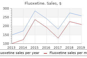
"Purchase fluoxetine with american express, womens health partnership."By: Peter Bartlett Bressler, MD - Associate Professor of Medicine
 https://medicine.duke.edu/faculty/peter-bartlett-bressler-md
Purchase 20 mg fluoxetine mastercardSpontaneous passage is likely if the calculi are small (less than three mm) and could additionally be aided by simple measures such as T-tube clamping. If the patient tolerates clamping and providing no untoward signs or issues develop, such a conservative strategy can be continued for a few weeks, on the end of which time the situation is reviewed radiologically. The varied methods out there for the non-surgical administration of retained stones are: (c) (d) �flushing �dissolution stone extraction through the T-tube tract �percutaneous �endoscopic sphincterotomy and stone extraction. The first three choices are relevant solely to sufferers with an indwelling T-tube whereas endoscopic stone extraction can be used in all sufferers. All the above methods are carried out beneath antibiotic cover due to the risk of cholangitis and septicaemia. Flushing is usually carried out with saline, heparinized saline or lidocaine�saline answer. Ductal calculi and cholangitis 723 Cholate infusion can dissolve ldl cholesterol stones however its efficacy is low and it has been changed by mono-octanoin, which acts extra rapidly and achieves full stone clearance in 40%. Percutaneous stone extraction via the T-tube tract was initially carried out by the Burhenne method using a Dormia basket introduced through a specifically designed steerable catheter to seize and extract the stone beneath fluoroscopic control. It has largely been changed by the versatile choledochoscopic approach which is successful in 90�95% of circumstances. A 4�6 week period of maturation of the T-tube tract is required earlier than the procedure may be performed safely. Thereafter, the T-tube tract is dilated to allow the introduction of the slim versatile choledochoscope. Endoscopic sphincterotomy with stone extraction is the most effective technique of coping with the issue of retained stones and may be carried out in sufferers with and without T-tubes. Surgical management of missed stones is reserved for these patients in whom the above methods have failed or complications have developed during or after tried endoscopic or percutaneous stone extraction. The administration of patients with recurrent ductal calculi is determined by their age and common condition. Endoscopic sphincterotomy and stone extraction is the first-line treatment and surgery (open or laparoscopic) reserved if this method fails. During surgery, the stones are removed atraumatically by the use of biliary balloon catheters, stone-grasping forceps or Dormia baskets as described beforehand. A completion verify via a choledochoscopic inspection or cholangiography abolishes or reduces the incidence of residual stones. In different conditions, recurrent ductal calculi are often multiple and related to gross dilatation of the bile duct and in some instances obvious distal ductal stenosis. This may be primary (papillary stenosis) or be secondary to trauma inflicted by steel bougies launched through the sphincter region on the time of exploration of the widespread bile duct. In sufferers with a quantity of ductal calculi, grossly dilated bile duct (>2 cm) or papillary stenosis, a drainage operation is indicated: choledochoduodenostomy or transduodenal sphincteroplasty. However, sphincteroplasty carries a major threat of pancreatitis and involves a sizeable duodenotomy. Recurrent ductal calculi Ductal calculi presenting 2 years or more after an operation are usually thought to be main. This discovering stresses the significance of avoiding non-absorbable material throughout operations on the biliary tract. The precise pathology stays unclear, but it appears probably that the clip is positioned too close to the frequent bile duct resulting in localized pressure necrosis. The internalized clip turns into coated with calcium bilirubinate to kind a brown pigment stone. The sufferers who Intrahepatic calculi (hepatolithiasis) Intrahepatic stones (hepatolithiasis) are prevalent in Southeast Asia but rare in Western countries. The distal finish is closed with a operating suture and the proximal finish is anastomosed in an endto-side style to the duodenum on the junction of the primary with the second part. Intrahepatic stones are categorized as: intrahepatic stones, which �primary related to intrahepaticform within the intrahepatic ducts and are duct strictures secondary, which type inside the extrahepatic ducts but migrate �subsequently to the intrahepatic ducts. Glucuronolactone deficiency could thus lead to elevated deconjugation of bilirubin diglucuronide with formation of calcium bilirubinate stones. The condition is also referred to as recurrent oriental cholangitis and has been lately reviewed. Epidemiology Primary intrahepatic stones are prevalent in East Asian international locations (Taiwan, China, Japan and Korea) and rare in Western nations, the Middle East and Africa. In Taiwan, primary ductal calculi account for greater than 50% of all cases of cholelithiasis and in Japan, 34%. Some have suggested that a low-protein food plan decreases glucuronolactone in bile, which is an Bacterial an infection of the biliary tree and bile stasis appear to be an important as the prevalence of micro organism within the bile of sufferers with hepatolithiasis exceeds 90%. All exhibit -glucuronidase enzyme exercise, which is believed to be answerable for the hydrolysis of the soluble bilirubin glucuronide to the water-insoluble unconjugated bilirubin which then combines with ionized calcium and precipitates, forming calcium bilirubinate stones. Intrahepatic stones are usually associated with strictures and dilatations of the intrahepatic ducts. In Taiwan, which has a very high incidence of intrahepatic lithiasis, infestation with C. Likewise for disease located solely in the best intrahepatic ducts, right anterior or posterior segmentectomy could additionally be necessary. Right hepatic lobectomy is however inadvisable due to surgical dangers especially as a result of the percutaneous cholangioscopic strategy provides a much safer and effective therapy. In the previous, an prolonged hepaticojejunostomy (Rouxen-Y) with a permanent cutaneous entry was used for the removing of stones from the best hepatic ductal system. However, this has been largely replaced by the percutaneous transhepatic treatment. When hepatolithiasis entails both intrahepatic ducts (more than 50% of patients), surgical treatment may be used for left lateral hepatic segmentectomy or left hepatic lobectomy and extended hepaticojejunostomy (Roux-en-Y) with everlasting cutaneous access. Some would nevertheless disagree with this and advocate percutaneous therapy for bilateral stones as the first possibility before recourse to surgical treatment. Percutaneous transhepatic approach the percutaneous transhepatic method has a quantity of advantages. This remedy is relevant to sufferers with multiple bilateral intrahepatic stones. Strictures can be dilated by balloons or by bougienage and the passage of Yamakawa catheters by way of the tract. However, percutaneous transhepatic cholangioscopic stone removal requires an experienced interventional endoscopist, preferably working with an interventional radiologist. The longterm results of percutaneous transhepatic cholangioscopy are much like those of surgery. The most important elements affecting the recurrence are the presence of strictures and bile stasis. Clinical features Many patients with intrahepatic stones may remain asymptomatic for many years and sometimes the situation is simply found throughout routine investigation.
Buy fluoxetine overnight deliveryIn the lower finish of the vertebral column, the dura mater narrows, and ends on the degree of the second sacral segment. It is prolonged at the filum of the dura mater toward the coccyx, and blends with the periosteum. The dura mater is separated from the partitions of the vertebral canal by the epidural space, containing semifluid fats and lots of veins. These veins Essential Anatomy and Function of the Spinal Cord a hundred twenty five are similar to the dural venous sinuses of the cranial cavity and lie between the periosteum of the vertebral canal and the dura mater. Prior to the extraction of the cerebrospinal fluid, stress measurements can be taken. A lumbar puncture can be taken to aid diagnosis of a variety of conditions including meningitis, subarachnoid hemorrhage or Guillan�Barr� syndrome (immunological condition inflicting injury to the myelin sheath). The lumbar puncture is carried out with the affected person lying on one facet with their legs pulled up toward their chest. The sample of cerebrospinal fluid is taken from beneath the extent of the second lumbar vertebra because the spinal wire terminates at that time. As the brain itself is insensitive, complications tend to be of a vascular origin, or of dural origin. It can be utilized during childbirth or trauma to the chest, stomach or pelvis in providing efficient pain administration (Faculty of Pain Medicine, 2010). It includes the insertion of a catheter into the epidural area applicable to the area to be provided with analgesia. It is crucial to follow local tips in its insertion, and monitoring of the affected person, and likewise to be conscious of any unwanted effects, and dangers associated with this procedure. There are eight in the cervical area, 12 in the thoracic region, 5 in the lumbar area, 5 within the sacral area and one coccygeal nerve. It is the dorsal root which carries information associated to afferent fibers and the ventral root carries efferent information. The nerve roots move for a small distance throughout the dural sac around the spinal twine. The roots then pierce by way of the dura and enter via the intervertebral foramina. At this point, the dorsal root ganglion can also be discovered and it has the cell bodies of the afferent fibers which are about to enter the spinal twine. Essential Anatomy and Function of the Spinal Cord 127 Distal to the ganglion, both the ventral and dorsal roots come together to form the frequent spinal nerve. The spinal cord, cylindrical in form as previously described, is bigger in the decrease cervical segments and within the lumbosacral territories. The lower cervical and first thoracic ranges are bigger where the brachial plexus arises from, to supply the higher limbs. At the decrease end, the lumbar and sacral areas are bigger due to the origins of the lumbosacral plexus innervating the lower limbs and pelvic organs. Just because the cranium protects the brain, the spinal twine is surrounded by the vertebral column, made up of a sequence of particular person vertebrae. Despite regional variations, typical vertebrae possess: (a) an enormous weight-bearing part the body, (b) a posterior arch which, with the back of the body, types the vertebral canal(or foramen) across the spinal twine; and (c) three processes which stand out from the vertebra. There is a large fibrous joint between the our bodies of adjacent vertebrae � the intervertebral disc � and smaller synovial joints exist between the transverse processes of adjoining vertebrae. In the thoracic area there are additional articulations for ribs, the primary and second cervical vertebrae are highly modified and the sacral vertebrae are fused collectively for power. The identical three layers of meninges that surround the brain additionally encompass the spinal wire. Again, the innermost layer, the pia mater intimately surrounds the spinal twine, whereas the outermost layer, the dura mater is partly hooked up to the bone. Since the spinal cord ends at L1/2, this is where the pia ends also (except for a thin projection, the filum terminale). Not solely are the meninges themselves protecting, but also the fluidfilled meningeal areas (such as the subarachnoid space) dampening down twine actions. It is enlarged within the cervical and in the lumbosacral regions, since these deal with the higher and decrease limbs, respectively. The spinal cord consists of a selection of segments (8 cervical, 12 thoracic, 5 lumbar and 5 sacral, plus 2�3 coccygeal). From each section, emerge two series of rootlets on each side; a dorsal series and a ventral series. These come together to kind a single dorsal root (on which is located a swelling, the dorsal root ganglion) and a single ventral root on all sides. The dorsal roots/rootlets are related to incoming (or afferent) sensory information similar to contact, ache, temperature, and proprioception. The ventral roots/ rootlets are associated with outgoing (or efferent) information, i. Soon after its formation, a spinal nerve divides into (1) a posterior primary ramus which supplies skin and muscle behind the vertebral column, and (2) an anterior main ramus which provides skin and muscle in front of the vertebral column. In the case of the nerves supplying the neck, upper limb and lower limb, the nerves come into shut proximity and intermingle as plexuses. Essential Anatomy and Function of the Spinal Cord 129 this is often interpreted as a safety device, since each muscle and (more importantly) each motion are actually innervated by a couple of spinal cord section. Nevertheless, lack of feeling (anesthesia) in a selected dermatome area may point out that there was damage or disease of the spinal wire segment (or spinal nerve/roots) supplying that area. Altered sensation over the world of distribution of a named peripheral nerve (rather than a dermatome) would counsel injury to a nerve distal to a plexus. This part is taken at the fifth lumbar vertebral level utilizing immunofluorescence on the left side for a neuronal marker NeuN. Based on detailed studies of neuronal soma measurement (revealed using the Nissl stain), Rexed (1952) proposed that the spinal grey matter is organized in the dorso-ventral axis into laminae and designated them into 10 groupings of neurons identified as I�X. The Input, Output and Functions of Each of the Laminae Within the Spinal Cord Lamina Characteristics I Small neurons and large marginal cells. Descending dorsolateral fasciculus fibers Some neurons projecting from the spinal twine (projection neurons), some passing to totally different laminae and some with axons confined to a lamina within the area of the dendritic tree of that cell. Nucleus accumbens and the lateral parabrachial area and reticular nuclei Pain, temperature and crude touch. Light mechanical stimulation Essential Anatomy and Function of the Spinal Cord 131 Table 7. The Input, Output and Functions of Each of the Laminae Within the Spinal Cord (cont. Triangular, fusiform and multipolar cell types discovered here Input Large main afferent collaterals. Descending fibers from the corticospinal and rubrospinal tracts Output Brainstem and thalamus via ipsilateral and contralateral spinothalamic tracts.
Purchase fluoxetine with american expressIt is a time period which indicates that the histopathologist is unable to come to a agency prognosis owing to the presence of options of both conditions. Ulcerative colitis 971 Radiology A plain belly radiograph is probably the most useful technique of identifying colonic dilatation. The instant barium enema is now not often used though it gives a superb report of the extent of the illness typically. The presence of intramural gasoline on the plain belly radiograph is a sign of imminent perforation and is subsequently a sign for instant surgical procedure. Unresponsiveness to medical therapy in acute colitis Failure to reply to medical treatment should be acknowledged early. The gastroenterologist and surgeon ought to confer at least every day to determine whether there has been enchancment, stagnation or deterioration. Deterioration regardless of enough medical remedy should be an indication for surgical procedure. Stagnation over a quantity of days with no sign of enchancment also needs to be an indication for operation. Clinical indicators that surgery is prone to be essential on the time of admission include a frequency of defecation of over 10 instances per 24 hours with the passage of blood at every defecation try, low albumin, low haemoglobin and a fall in lean body mass of more than 10%. Previous acute assaults, poor basic well being and social circumstances affected by the illness must be taken into account. A significant historical past of chronic illness and social incapacity ought to sway the choice in favour of surgery. The former is intended to induce a remission,the latter to preserve a remission as quickly as achieved. Both could be given as suppositories or as an enema, the selection relying on the proximal extent of disease. They are formulated to shield the aspirin from degradation earlier than it arrives in the colon. Proctitis refractory to this therapy may respond to different preparations, including bismuth, nicotine and witchhazel. Treatment Best care is likely to be achieved by a multidisciplinary team including medical employees, specialist nurses, nutritionists and stomatherapists with social and psychological support. Collaboration between gastroenterologist and surgeon is important and should include patient sharing the place appropriate, joint outpatient consultations for difficult instances, and early involvement of the surgeon in acute disease. Medical treatment Medical remedy entails bed relaxation and the correction of water and electrolyte depletion by intravenous infusion. Intravenous vitamin may be indicated in severely malnourished sufferers, as judged by a lower in lean body mass and serum albumin. Intravenous prednisolone (60 mg daily) and an H2-receptor antagonist or a proton pump inhibitor to defend against upper gastrointestinal ulceration are given. Ciclosporin has been reported to induce remission in over 50% of sufferers unresponsive to steroids but early relapse could occur, resulting in the same scientific situation inside a brief interval. More lately, a quantity of research have reported using organic brokers for the therapy of acute ulcerative colitis. In a examine of 30 patients with active ulcerative colitis treated with infliximab between 2000 and 2006 at Oxford, 53% of patients came to colectomy at a median time of one hundred forty days after their first infusion (range 4�607). The position of immunotherapy in the acute setting remains to be established in a selection of randomized controlled trials. Surgery is absolutely indicated in circumstances with acute toxic dilatation or perforation. This ought to document each defecation with an evaluation of volume and consistency of stool and the presence or absence of blood on each event. It may be silent in a affected person on large doses of steroids, and will become evident only by the presence of free gasoline on the plain stomach radiograph. Medical treatment of persistent proctocolitis Medical administration consists of anti-inflammatory, dietary, symptomatic and psychological remedies. Prednisolone is given in an initial dose of 40 mg, steadily lowering this as remission occurs over the next few weeks. Patients may have experienced a quantity of hospitalizations, durations off work, disruption of household life and schooling and other social results of continual illness. Steroid dependence A response to steroids could additionally be maintained solely by continuing the remedy with relapse on withdrawal. If various medicine corresponding to immunosuppression is unsuccessful, then surgical procedure is indicated except there are specific causes towards. Recurrent acute exacerbations the choice for surgical procedure will depend on the frequency and severity of attacks. Surgery during an acute assault will usually take the type of a colectomy with ileostomy. A decision taken during remission could allow an elective definitive process corresponding to restorative proctocolectomy to be carried out as the first stage process. Severe signs the patient could additionally be systemically well however severely inconvenienced by frequency and urgency of defecation significantly if related to urge incontinence. However, the activityrelated polyarthropathy does reply, as will some instances of pyoderma gangrenosum, although the latter could enhance only slowly over several months. Antidiarrhoeal brokers together with codeine phosphate and loperamide are usually effective in reducing frequency and urgency. Lomotil (atropine and diphenoxylate) is sometimes efficient the place there has been a poor response to the others. Bone densitometry must be carried out where steroid medicine has been prolonged. Unresponsiveness to medical treatment in persistent colitis and surgical indications Most sufferers requiring surgery have extensive disease. Very often, a affected person with distal illness may require surgery, normally because of severe native signs. The indications for elective surgery embrace: therapy �unresponsiveness to medical young retardation of growth within the �malignant transformation. Steroid medicine itself leads to early fusion of epiphyses, leading to permanent stunting of development. Patients in this category are usually beneath the care of a paediatric skilled within the assessment of progress. However, throughout the years of puberty a delay in surgical procedure may happen, partly due to the antipathy of the paediatrician and/or patient (or parents) to an ileostomy. Malignant transformation the presence of high- or low-grade dysplasia or an established invasive tumour is an indication for surgery. The surgical method ought to be as if invasion had occurred since this can be determined solely by examination of the resected specimen. Surgical therapy Acute colitis (emergency surgery) Surgery has a major position in the management of acute colitis. The want for surgery is best during the first 12 months after onset of the illness.
Purchase fluoxetine discountThe endoscopist then performs a sphincterotomy and stone extraction with the patient within the supine place. The reported experience with the laparoendoscopic technique though restricted has been totally beneficial, however the approach requires further resource and the supply of an skilled versatile endoscopist. The perceived risk of acute postoperative pancreatitis with this system has not materialized in any of the reported series. Direct supraduodenal frequent bile duct exploration that is indicated for large or occluding stones. The dissection is minimal and consists of exposure of the anterior wall of the common bile duct after downward displacement of the duodenum. The dimension of the choledochotomy must be roughly half to three-quarters the maximum diameter of the biggest stone. Avoidance of a large choledochotomy reduces the amount of intracorporeal suturing needed, and, in view of the elasticity of the frequent bile duct, the opening may be stretched to accommodate giant stones. Ductal calculi and cholangitis 721 (a) (b) (c) facet, balloon dislodgement and direct visual wire basket extraction after the insertion of a 4 mm versatile choledochoscope. Primary closure of the choledochotomy is carried out, throughout which the frequent bile duct is continually irrigated with saline by way of the cystic duct drainage cannula. In both case, a completion cholangiogram is crucial to ensure ductal clearance and a subhepatic drain is important. The cystic duct drainage cannula or the T-tube is firmly anchored to the abdominal wall and related to a biliary drainage bag. When a cystic duct drainage cannula is used, the postoperative cholangiogram is carried out on the second day; if this is normal, the cannula is sealed with a Luer lock and coated with an occlusive dressing. In the absence of any bile leakage from the subhepatic drain over the next 24 hours, the drain is removed and the patient discharged the following day with the cystic duct drainage cannula in situ. In the aged, diabetics and patients on immunosuppressive medication, the T-tube is saved in place for at least 2 weeks as the maturation of the T-tube tract is impaired in these patients. They are normally small floating calculi with out dilatation of the frequent bile duct. This expectant policy will incur a particular morbidity from jaundice, cholangitis or pancreatitis and some (albeit few) sufferers will die on account of one of these issues. Routine completion choledochoscopy/cholangiography virtually abolishes this complication. Retained ductal calculi following biliary tract surgery are both identified within the quick postoperative interval by the postoperative T-tube cholangiogram or present with recurrent symptoms usually inside 2 years of cholecystectomy with out exploration of the frequent bile duct. Ductal stones presenting past this interval are usually thought-about to be of the primary selection. Certain basic concerns apply with regard to the administration of patients with residual calculi following biliary tract surgical procedure. Symptomatic sufferers can present with upper stomach pain, occasional fever, rigors and, less frequently, jaundice. In addition, these patients have moderate elevations of serum transaminases and delicate to average iron-deficiency anaemia. Untreated sufferers with longstanding illness with recurrent attacks of bacterial cholangitis can develop biliary cirrhosis with coagulation defects, low serum albumin and the event of ascites. Ultrasound scanning is used for screening only as its diagnostic accuracy for documenting the full extent of hepatolithiasis is limited. Cholangitis Acute bacterial cholangitis is a serious, life-threatening emergency attributable to infection of an obstructed biliary tract. The systemic manifestations of the acute sickness end result from bacteraemia secondary to cholangiovenous reflux induced by the biliary hypertension (<20 cmH2O). The most typical obstructing agent is an occluding stone within the widespread bile duct, adopted by bile duct strictures (including sclerosing cholangitis) and tumours of the bile ducts, pancreatic head and periampullary lesions. Less commonly, cholangitis is secondary to bilioenteric anastomoses, spontaneous bilioenteric fistulas, cystic disease of the biliary tract and duodenal diverticula. The risk components for cholangitis following this investigation are the presence of fever before the process and malignant biliary obstruction. Cholangitis brought on by contamination Treatment Currently, two major therapy modalities are used with particular indications: �surgical operative administration �percutaneous therapy. However, percutaneous transhepatic cholangioscopy removal offers good results and is rising because the preliminary therapy in patients with bilateral illness. Surgical remedy this goals at each full removal of stones and dilation of bile duct strictures to ensure enough drainage with no bile stasis, important for the prevention of recurrence and bacterial infections. At operation gross purulent cholangitis was observed because the biliary tract was transected above the tumour. Biliary decompression could also be completed surgically, endoscopically or by percutaneous methods. For most sufferers with cholangitis and ductal calculi, endoscopic decompression by sphincterotomy and extraction of calculi is usually favoured, especially if the affected person is elderly. The alternative is surgical exploration with ductal clearance and insertion of a T-tube. Cholangitis secondary to biliary instrumentation usually requires operative remedy. Percutaneous transhepatic drainage is helpful in patients with cholangitis complicating strictures and malignant obstruction. The general reported mortality of patients requiring urgent decompression for extreme cholangitis is 15�20%. Sclerosing cholangitis that is an obscure dysfunction of unsure aetiology, which outcomes in a progressive fibrous obliteration of the biliary tract. The distinction between major and secondary varieties is now not held to be valid. The term major was formerly used to indicate no previous biliary surgery or biliary tract disease. Often, however, sclerosing cholangitis happens as a secondary complication of inflammatory bowel illness, normally ulcerative colitis and, much much less commonly, Crohn illness. The situation is presently considered an immune-complex dysfunction evoked by endotoxin�antibody complexes which were recognized in the peripheral blood of patients with inflammatory bowel disease. The classification of the dysfunction is predicated on the extent of involvement of the biliary tree by the fibrous obliterative process Table 25. The gross fibrous thickening ends in localized or a number of stricture formation. Although the ductal epithelium is regularly regular, it could turn out to be ulcerated and exhibit saccule formation. In Asia, recurrent pyogenic cholangitis is a frequent cause of recurrent bacterial cholangitis and is known as recurrent oriental cholangitis. Aside from toxicity, the excessive intermittent pyrexia is accompanied by extreme rigors.
Buy fluoxetine 10 mg without prescriptionSolitary rectal ulcer the time period solitary rectal ulcer is strictly a misnomer, as the lesions may be a quantity of and ulceration could not necessarily be current. The macroscopic look is of a pink thickened space of rectal mucosa often with a shallow ulcer within the centre. Clinically, all patients have difficulty in defecation which includes going to the bathroom a number of instances a day but only truly defecating once or twice. If a inflexible Aganglionosis and/or myenteric plexus lesions Other than drugs, constipation may be caused by malfunction of the intrinsic nervous system of the massive intestine and this in flip could be subdivided into Hirschsprung disease and idiopathic megacolon and megarectum. These are dealt with as separate points in the following sections of this chapter. Constipation because of extracolonic factors Extracolonic elements that may give rise to constipation include: �illness inflicting immobility. However, the only accurate means of demonstrating an intussusception is by using defecating proctography. Treatment could be tough and a conservative strategy must be taken in the first instance. A high-fibre food regimen and using suppositories might forestall excessive straining and alleviate the symptoms. Complications the main complication of pseudo-obstruction is faecal peritonitis secondary to perforation of the caecum. Treatment In the first occasion, administration consists of intravenous fluids, nasogastric aspiration and decompression with a flatus tube inserted at rigid sigmoidoscopy. Concomitantly, you will want to appropriate any metabolic disturbances, deal with infections and cease any medications that will affect colonic motility. This reduces the diameter of the caecum in about 70% of cases, but in about 40% repeated colonoscopy shall be required. Recurrence of the pseudo-obstruction may be decreased by inserting a drainage tube into the right facet of the colon on the time of the first colonoscopy. Surgery for acute colonic pseudo-obstruction is indicated if all these conservative measures fail to convey about a discount within the size of the caecum. In the absence of indicators suggesting ischaemia or perforation of the bowel, the operation of selection is a tube caecostomy via a limited proper iliac fossa incision to expose the caecum. A large Foley catheter can then be used to intubate the caecum and this ought to be retained for about 3 weeks. However, if there are signs of ischaemia or perforation, a midline laparotomy ought to be used. Under most circumstances, a direct anastomosis ought to be deferred and an ileostomy and mucus fistula common. It ought to be remembered that colonic pseudo-obstruction is associated with a big mortality, largely owing to the underlying sicknesses which are associated with this situation. Colonic pseudo-obstruction Acute colonic pseudo-obstruction or Ogilvie syndrome is characterized by marked dilation of the colon in the absence of mechanical obstruction. It nearly all the time happens in hospitalized sufferers and the vast majority have an related condition corresponding to an infection, widespread malignancy, recent surgical procedure or trauma particularly to the spine. Clinical features the medical features of acute colonic pseudo-obstruction carefully mimic acute large bowel obstruction. The affected person typically has colicky belly pain and progressive distension of the abdomen is the rule. Constipation is common, though not absolute, as some sufferers will proceed to pass a small amount of flatus or liquid stool. Diagnosis the analysis is mostly made on plain abdominal radiograph, which shows the everyday appearance of a distal colonic obstruction often with the cut-off within the descending colon. Measurement of the diameter of the caecum on the radiograph is necessary as perforation is widespread as soon as this exceeds 12 cm. It may be defined as stomach discomfort or pain associated with defecation or a change in bowel behavior and with an element of disordered defecation. They consist of an abnormal stool frequency (more than three times a day or less than three times a week), irregular stool consistency (hard, loose or watery), abnormal passage of stool (urgency, straining or tenesmus), the passage of mucus and belly bloating or distension. The disease presents variably, starting from mildly symptomatic fluctuation in bowel behavior and left decrease quadrant belly pain, by way of haemorrhage and localized sepsis to colonic fistulation, perforation and life-threatening peritonitis. In addition, signs might current as an isolated occurrence, or develop right into a persistent complaint. Considered a disease of Western society and of aged sufferers, its incidence is rising along with related episodes of hospitalization. Despite the numerous associated healthcare burden, the aetiology, pure historical past and optimum management of this disease remains controversial. The terminology applied to describe the totally different medical displays and disease stages related to diverticular illness can be variable. The term mychosis is used in the context of diverticular illness to describe muscular shortening and thickening of the colonic wall, commonly seen at surgery. The first famous reference to colonic diverticula as a pathological entity was made by the French surgeon Alexis Littre within the 1700s, though this was not in relation to diverticular illness per se. The pathology we recognize right now as diverticular disease was first described by Jean Cruvehiler in 1849. Its absence from early surgical textbooks presently suggests the illness was not as prevalent, or not as acknowledged, as at present. Mayo reported the first surgical resection for sophisticated diverticulitis in 1907, advocating major resection, though the illness remained uncommon. Although this case collection promoted primary resection, staged resection consisting of drainage and stoma, interval resection and subsequent stoma reversal remained routine till the development of other therapy paradigms within the fashionable era. Similarly, elective surgical procedure for recurrent disease has seen a step-change in latest times. Normally it will take the type of barium enema and sigmoidoscopy or colonoscopy. Certainly, if a patient has irritable bowel-type signs associated with rectal bleeding, weight loss or different suspicious symptoms, they need to be totally investigated. The prognosis, however, rests on medical options and the diagnostic criteria agreed on the multinational consensus meeting on functional gastrointestinal issues held in Rome in 1999 are as follows. If constipation is a major function then dietary fibre should be increased by using wheat, bran or bulking brokers. For stomach ache, anticholinergic or antispasmodic brokers similar to mebeverine could additionally be of value. It is characterised by episodes of severe anal or lower rectal pain which last for a variable length of time, starting from a couple of seconds as much as several minutes. The cause of the pain might be clean muscle spasm but the underlying cause is obscure. The authentic findings of this research have been challenged in recent years, and current research suggests a extra benign course of recurrent disease. This, along with the not insignificant morbidity related to elective resection, signifies that a number of of the commonly accepted remedy algorithms for diverticular illness are presently in the method of being redefined.
Fluoxetine 20 mg with amexThe diameter of the transduodenal section is often 5 mm and that of the main duodenal papilla varies from zero. The commonest web site for calculus arrest or impaction is simply proximal to the transduodenal phase. The bile and pancreatic ducts are illustrated splayed aside to facilitate the demonstration. Its appearance might vary from the identical old well-defined papilla with various degrees of projection to a flattened despair between the mucosal folds. Irrespective of its exact configuration, the most important duodenal papilla frequently has a dorsal mucosal fold. The minor (accessory) papilla is more proximally situated and assumes scientific importance only in sufferers with pancreas divisum. The exercise of the choledochal sphincteric complicated is unbiased of the duodenal musculature but may be influenced by it. Thus, the impact of certain medicine on the choledochal sphincter differs from their action on the duodenal wall, and duodenal muscular peristaltic exercise has no vital effect on the common bile duct strain. Contraction of the longitudinal muscle tends to open the duct lumen, whereas the circular muscle has the opposite effect. These contracted (systolic) and relaxed (diastolic) states of the choledochal sphincter lead to fairly distinct appearances of the lower finish of the common bile duct at cholangiography. The cystic lymph node is mostly situated on the junction of the cystic with the common hepatic artery. The cystohepatic triangle is virtually obliterated within the presence of Mirizzi syndrome (see below). The hepatic capsular lymphatics drain into the thoracic duct except these on the superior floor of the liver, move from which reaches the retrosternal lymph nodes through several channels. Distally, the gallbladder lymphatics and people of the extrahepatic bile ducts drain into the cystic lymph node, which is situated near the cystic artery close to its origin from the proper hepatic artery, and to different nodes lateral to the decrease finish of the bile duct, notably the retroduodenal segment. The distal lymphatic drainage of the gallbladder follows three routes: (1) cholecystoretropancreatic lymphatics, which run downwards anterior and posterior to the common bile duct to finish within the retroportal nodes on the posterior floor of the head of the pancreas; (2) cholecystocoeliac lymphatics, which run to the left by way of the hepatoduodenal ligament and drain in the coeliac nodes; and (3) the cholecystomesenteric lymphatics, which run to the left in front of the portal vein and drain in lymph nodes at the root of the superior mesenteric vessels. The cholecystoretropancreatic drainage is considered the primary pathway with respect to nodal spread from cancer of the gallbladder. Cystohepatic triangle of Calot the cystohepatic triangle is important in biliary surgery particularly within the performance of cholecystectomy. It is a triangular fold of peritoneum containing the cystic duct, cystic artery, cystic node and a variable quantity of fat. It also often contains the proper hepatic artery, which often enters the triangle behind the widespread hepatic duct and before it offers off the cystic artery, (a) (b) Hepatic artery the grownup hepatic artery is derived from the middle of the three primordial arteries that supply the fetal liver. The usual association is for the common hepatic artery to come up from the coeliac axis. It incorporates the cystic artery and lymph node and the right hepatic artery as it emerges from behind the frequent hepatic duct. The vast majority of aberrant/anomalous bile ducts come up from the proper ductal system (especially the dorsocaudal branch of the best hepatic) and 80% are situated in the cystohepatic triangle of Calot. Surgical biliary physiology 677 arteries behind the antroduodenal region, it arches upwards along the left side of the bile duct and in entrance of the portal vein. It then bifurcates into the proper and left hepatic arteries normally fairly close to the liver. The right hepatic artery normally crosses behind (rarely in front of) the frequent hepatic duct before giving rise to the cystic artery. Low division of the hepatic artery is encountered in 15% of sufferers when the right hepatic artery programs behind the portal vein. The essential anomalies are the outcomes of persistence of the left or proper primordial hepatic arteries. This anomalous vessel then arises from the left gastric artery or instantly from the aorta and traverses the lesser omentum to enter the liver within the umbilical fissure. Persistence of the best primordial artery ends in an anomalous right hepatic artery originating from the superior mesenteric. It ascends to the liver behind the pancreas and duodenum to reach the free edge of the hepatoduodenal ligament. Again it is very uncommon for a persistent right primordial hepatic artery to present the only arterial blood provide to the liver. Knowledge of abnormal portal vein anatomy is important in surgical resections of the liver, hepatic transplantation and many percutaneous interventional procedures, together with transhepatic portal vein embolization to induce contralateral liver hypertrophy in patients, transjugular intrahepatic shunt, etc. Thus for example, failure to recognize a Z-type portal vein variant throughout a left liver resection or when harvesting a residing donor liver transplant may result in loss of perfusion to the right anterior sector. Trifurcation of the portal vein might require two separate anastomoses when the proper liver is transplanted. The portal vein then courses upwards behind the superior part of the duodenum and ascends to the liver in the best border of the lesser omentum behind the frequent bile duct on the proper and hepatic artery on the left to the right extremity of the porta hepatis, where it bifurcates into the best and left portal veins which accompany the corresponding branches of the hepatic artery into the hepatic parenchyma. The portal vein bifurcation may be intrahepatic or extrahepatic (roughly equal incidence). The portal vein is surrounded by the hepatic plexus of nerves and numerous lymphatic channels and some lymph nodes. The left portal vein provides branches to the caudate lobe before it enters the liver. Surgical biliary physiology Hepatic ldl cholesterol and bile acid physiology the liver plays a key function within the metabolism of ldl cholesterol. It regulates the uptake of dietary cholesterol, its de novo synthesis and the excretion of this lipid in bile both as free ldl cholesterol or as bile acids. There is growing evidence for metabolic compartmentalization of ldl cholesterol inside the hepatocyte, i. The major bile acids are conjugated throughout the hepatocyte with the amino acids glycine and taurine, before being secreted into the bile canaliculi. Toxic relatively insoluble secondary bile salts (reabsorbed from the small intestinal pool) similar to lithocholic acid are sulphated previous to excretion. Cholesterol is transported into the bile canaliculi as phospholipid�cholesterol vesicles. The formation of phospholipid�cholesterol�bile acid micelles is a postcanalicular event induced by the excessive concentrations of bile acids in the hepatic bile. There is evidence that high concentrations of deoxycholic acid could promote the hepatic secretion of cholesterol saturated bile and thereby induce the nucleation of cholesterol crystals. Variant anatomy of portal vein Many anatomical variants of the portal vein have been described, including duplications, congenital absence and absence of portal vein branching (single portal vein enters the best liver and courses into the left, giving only segmental branches), however all these are rare. There is a small bile acid-independent fraction of bile secretion, which is lowered in experimental cholestasis, whereas the bile acid-dependent fraction increases beneath these conditions. Somatostatin decreases each bile acid circulate and bile ductular secretion, indicating a suppression of de novo bile acid synthesis by the hepatocytes.
Buy fluoxetine with mastercardThey are then coughed up to be swallowed into the gut where they mature as grownup worms (up to 40 cm in length) within the jejunal lumen. This is adopted by higher gastrointestinal endoscopy and colonoscopy for inspection of the mucosa and for procurement of biopsy material. Biopsies are cultured for cytomegalovirus and mycobacteria, adenovirus and herpes simplex virus. Clinical features Initially, the signs are brought on by the larval migration especially to the lungs (wheezing, fever, cough, chest pain and dyspnoea) including established asthmatic attacks. The symptoms attributable to the grownup worms include belly ache with colicky assaults, nausea, vomiting, anorexia, weight reduction, diarrhoea, malabsorption and anal itching. Treatment If a selected an infection is established remedy is directed to eradicating the organism accountable. Patients will benefit from supportive therapy with rehydration and electrolyte supplements. Plasma or serum citrulline assays have been launched as an accurate assessment of the enterocyte cell mass. Hypoalbuminaemia is frequent and the liver operate exams may reveal evidence of liver harm. Symptoms are usually present in sufferers with longstanding infestations with high worm loads. They embrace asthenia, tiredness, weight reduction and weak spot in affiliation with iron-deficiency anaemia. To some extent, the range and severity of signs vary extensively and depend upon the dietary standing and overall basic health of the affected person, the species of worm and the extent of infestation. Treatment Eradication therapy is normally with one of many benzimidazole compounds similar to mebendazole or albendazole or piperazine. At operation an try should be made to manipulate the mass of tangled worms into the caecum. Occasionally, a resection may be essential if the viability of the bowel is in any doubt or if a perforation has occurred. Mebendazole has a remedy price of 76�95% and is used because the first-line remedy of selection for adults and youngsters over the age of 2. In some areas where mebendazole has been used for long intervals, resistance to the drug has been reported. Piperazine is utilized in kids (between three months and a pair of years) but can be used as an alternative to mebendazole in adults normally combined with a purgative (senna) to guarantee full expulsion. Levamisole (nicotinic acid antagonist) is very effective and is given as a single dose. Diagnosis the diagnosis is made by the identification of ova in the faeces or grownup worms in duodenal or jejunal fluid. Treatment the first stage of remedy is the correction of anaemia utilizing either oral or injectable iron. Benzimidazole compounds, mebendazole or albendazole or pyrantel pamoate are used to clear the infestation. Small bowel circumstances inflicting malabsorption Small bowel bacterial overgrowth In this syndrome, also referred to as dysbiosis, the small gut becomes colonized by micro organism with a rise in the focus of organisms that are normally confined to the lower small bowel and the colon. This is usually caused by surgery or disease which results in extra micro organism getting into the small intestine, or from delayed clearance of bacteria due to stasis (stagnant or blind loop syndrome). The larvae enter the physique by penetrating into pores and skin, usually the only of the foot, forming an itchy lesion. The grownup worm lives hooked up by hooks to the mucosa of the jejunum and ova are excreted within the faeces. However, ancylostomiasis can produce minimal signs if the infestation is In some circumstances bacterial overgrowth might develop within the absence of an apparent native cause significantly in sufferers with Small bowel circumstances causing malabsorption 917 malnutrition or immune deficiency. This along with the action of the dehydroxylated bile salts, enterotoxin and osmotic load created by the fermentation of the major dietary parts accounts for the diarrhoea which is seen in these patients. Bacterial colonization results in intestinal mucosal damage characterised by patchy inflammatory adjustments within the lamina propria that are accompanied by alterations in the concentrations of the brush border enzymes. The increased bacterial inhabitants deconjugates the intraluminal bile salts by removing the glycine or taurine moiety. In addition the micro organism dehydroxylate the steroid nucleus on the C7 position with the formation of deoxycholate and lithocholate which tend to precipitate on the intraluminal pH levels of the small gut. This leads to a web reduction of the focus of effective bile salts in the lumen of the small bowel to ranges below that required to kind micelles. The bacterial species that are answerable for bile salt deconjugation are eubacteria, bacteroides and corynebacteria. These micro organism additionally bind vitamin B12 and convert it to inactive derivatives (cobamides) which block the ileal receptors for the vitamin in addition to for intrinsic factor. Often, nevertheless, the anaemia has a dimorphic picture due to an iron-deficiency component because of chronic blood loss from the primary lesion itself or from malabsorption of iron. Folate deficiency is uncommon as the micro organism synthesize folate in substantial quantities and some patients might actually exhibit a high serum folate. There is some malabsorption of carbohydrates and proteins though that is hardly ever significant. The primary purpose for malnutrition in sufferers with bacterial overgrowth is a diminished dietary intake, which accounts for progress retardation in children. The micro organism also metabolize triglycerides to free fatty acids which they hydoxylate to kind hydroxy fatty acids. These impair the absorption of water in sodium by the intestinal mucosa of each the (a) Clinical options the symptoms of bacterial overgrowth are varied. Initially the sufferers could exhibit signs referable to the underlying pathology. The signs and signs of bacterial overgrowth itself are sometimes non-specific and embrace malaise, nausea and vomiting, extreme borborygmi and weight reduction. Diarrhoea is extraordinarily frequent and is normally watery but frank steatorrhoea with cumbersome pale offensive stools which are difficult to flush is less frequent. Diagnosis A full blood count is essential to assess the extent and kind of anaemia which is often present in these patients. Estimations of urea and electrolytes, albumen and complete protein ranges are also necessary. The most useful take a look at for assessing the presence of bacterial overgrowth, small bowel nonetheless, is the hydrogen breath check (see section small bowel investigations). Complications the problems of bacterial overgrowth include glossitis, stomatitis, anaemia, hypoproteinaemia with peripheral oedema, tetany, osteomalacia and rickets and progress retardation in kids. Occasionally neurological manifestations similar to paraesthesiae and peripheral neuropathy may be present in affiliation with vitamin B12 deficiency. Treatment Surgical remedy of the underlying conditions wherever potential is the definitive and curative treatment. As control of the anaerobic organisms seems to be probably the most useful goal, metronidazole is generally preferred, though tetracycline can additionally be of worth. A course of antibiotics is often administered for about 2 weeks at a time and though repopulation of the gut by bacteria occurs soon after discontinuation of the remedy, symptomatic enchancment might final for several months. This is believed to work by restricting the vitamin obtainable for micro organism within the higher gastrointestinal tract and reduces the quantities of alcohol and natural acids produced by bacterial fermentation.
Cheap 10mg fluoxetine fast deliveryEarly stories demonstrated an elevated incidence of symptomatic gallstones in females on the oral contraceptive pill, the relative risk being estimated at 2. A lithogenic index of unity or greater signifies that the bile is supersaturated with respect to cholesterol. The regular ratio is focus bile salts + focus phospholipids/concentration ldl cholesterol = 10:1. The source of the supersaturated bile is the liver, from either increased synthesis of ldl cholesterol or a decreased synthesis/ secretion of bile salts and phospholipids. Most non-obese sufferers with cholesterol gallstones have a decreased absorption as the cause of the diminished bile salt pool in these sufferers. The most likely explanation of the smaller pool (and supersaturated bile) is an exaggeration of the normal feedback mechanism between the bile salt return and the hepatic synthesis of bile acids together with an increased enterohepatic cycling possibly consequent on abnormal excessive gallbladder contractility. Although gallstones develop solely in individuals with supersaturated bile, many patients with this abnormality never develop gallstones. Recently a protein with marked nucleation promotion exercise for cholesterol crystals that binds to concavalin A has been isolated from the bile of sufferers with and with out gallstones. Biliary sludge Biliary sludge consists of mucin, calcium, monoconjugated bilirubin and ldl cholesterol and is now thought to be the direct precursor of gallstones. A number of stories have documented symptoms together with biliary-type ache in sufferers who were discovered to have biliary sludge however no gallstones. Other components There is little doubt that the above account presents an incomplete image of the pathogenesis of ldl cholesterol stones. The position of calcium is indicated by the presence of calcium salts in the majority of stones, and the experimental demonstration in the prairie canine model that ldl cholesterol vesicle aggregation and the induction of ldl cholesterol gallstones is related to an increase within the whole and free ionized calcium concentrations in the gallbladder bile. That the gallbladder plays an essential role in gallstone formation (both cholesterol and black pigment) is obvious by the reality that 85�90% of stones are encountered on this organ somewhat than within the bile ducts. Biliary vesicles Under regular situations, the cholesterol�phospholipid vesicles are comparatively stable and disappear as micelles form and take up their cholesterol and phospholipid constituents. In the pathological state, the part change to micelles is incomplete and as more phospholipid than ldl cholesterol is extracted into the micellar aggregates with bile salts, biliary vesicles with an abnormally high cholesterol�phospholipid ratio are produced. These abnormal vesicles are unstable and are the supply of cholesterol monohydrate crystals in the bile. The crystallization is preceded by aggregation and fusion of the excessive cholesterol-containing vesicles. Kinetic balance between nucleating and antinucleating elements Bile incorporates substances which both inhibit (apolipoprotein A1) or promote (mucin) the expansion of cholesterol crystals. Thus the event of cholesterol gallstones is influenced by the stability between these two kinetic elements. The biliary proteins include enzymes, transport proteins, hormones, plasma proteins and mucin. The proof for the important role of mucin in the nucleation and growth of cholesterol crystals is overwhelming. Hypersecretion of densely glycosylated mucin is observed in experimental fashions of gallstone formation; mucin is a major part of biliary sludge and stone formation seems to begin in the mucous gel. Thus gallbladder mucus is thought to be concerned each as a nucleation-promoting agent and in providing the right milieu Black pigment stones Much much less is known in regards to the pathophysiology of black pigment stones. The hypothesis that sufferers with black pigment stones have a bile which is supersaturated with calcium bilirubinate has had some experimental backing in a dietary model of pigment stones by which a major increase in the gallbladder concentrations of unconjugated bilirubin and calcium have been documented. Calcium is a common part of black pigment stones and each free and whole ionized calcium is increased in canine models of pigment gallstone illness. As the hepatic bile concentration is unchanged, the altered chemical composition is thought to be secondary to altered gallbladder perform. Patients with bile acid malabsorption (after ileal resection) have low biliary ldl cholesterol in addition to a lowered bile salt pool. They exhibit an elevated threat of gallstone formation, predominantly of the black pigment kind. This statement has led to the hypothesis that biliary ldl cholesterol may exert a protecting motion on the gallbladder mucosa and, accordingly, low biliary cholesterol renders the mucosa vulnerable to direct Gallstones 707 Brown pigment stones these ductal calculi are attributable to an infection by Gram-negative micro organism corresponding to E. In many Eastern international locations the place infestation with Ascaris lumbricoides is endemic, the eggs of this parasite have been repeatedly recognized within the nucleus of brown pigment stones, however the trigger is the associated bacterial an infection of the biliary tract. Likewise, the formation of each intrahepatic and extrahepatic ductal calculi in recurrent pyogenic cholangitis is because of infection of the biliary tract by Gram-negative micro organism. The bacterial -glucuronidase is implicated within the hydrolysis of conjugated bilirubin with consequent precipitation of insoluble calcium bilirubinate. Brown pigment stones are encountered in biliary tract conditions related to stasis and infection such as persistent obstructive illness, indwelling biliary endoprostheses and around non-absorbable suture materials or metallic clips utilized in biliary tract surgical procedure. Clinical syndromes of gallstone illness the symptomatology of gallstone illness is varied. Often non-specific, the signs could additionally be acute, persistent or totally absent when gallstones are identified as an incidental discovering through the investigation of patients for unrelated issues. The differentiation between silent and symptomatic gallstones is important since this affects management in the individual case. This is particularly necessary within the number of patients for elective cholecystectomy. The factors inside the gallbladder which favour precipitation of ldl cholesterol crystals and the growth of ldl cholesterol microliths into discernible stones are outlined in the text. Symptomless (silent) gallstones Most surveys have shown that silent gallstones heavily outnumber the symptomatic ones. Silent gallstones are diagnosed as incidental findings most commonly by abdominal radiographs. Another argument for cholecystectomy in the past has been the prevention of gallbladder most cancers, the development of which is known to be associated with the presence of gallstones. However, carcinoma of the gallbladder is rare and the general operative mortality with the widespread adoption of prophylactic cholecystectomy in patients with silent gallstones would definitely exceed that as a result of most cancers of the gallbladder by a significant margin. The first pertains to patients undergoing surgical intervention for different situations in which gallstones are found at laparotomy. In one report, 50% of these patients developed problems or symptomatic illness subsequently and 12% required cholecystectomy within 30 days after the operation. On the opposite hand, concomitant cholecystectomy increases the postoperative morbidity. The second group which deserves cholecystectomy for asymptomatic gallstones is made up of acromegalic sufferers on long-term therapy with somatostatin analogue, which regularly produces giant gallstones. Patients with a calcified gallbladder have a significant threat of growing most cancers of the gallbladder and for this reason cholecystectomy is indicated even when the condition is asymptomatic. Symptoms and signs Most generally, patients with continual cholecystitis complain of recurrent attacks of epigastric or right hypochondrial ache, usually radiating to the right facet of the again and, much less generally, to the shoulder blade.
References - Ramani A, Desai M, Steinberg A, et al: Complications of laparoscopic partial nephrectomy in 200 cases, J Urol 173:42-47, 2005.
- Gaughan EM, Dezube BJ, Aboulafia D, et al: Human immunodeficiency virusnassociated renal cell carcinoma: a transatlantic case series, Clin Genitourin Cancer 6(2):86n90, 2008.
- Shui IM, Mondul AM, Lindstrom S, et al; Breast and Prostate Cancer Cohort Consortium Group: Circulating vitamin D, vitamin D-related genetic variation, and risk of fatal prostate cancer in the National Cancer Institute Breast and Prostate Cancer Cohort Consortium, Cancer 121(12):1949n1956, 2015.
- Bade JJ, Laseur M, Nieuwenburg A, et al: A placebo-controlled study of intravesical pentosanpolysulphate for the treatment of interstitial cystitis, Br J Urol 79(2):168n171, 1997.
- Palmer LS: Evaluation and targeted therapy of voiding dysfunction in children, Urology 92(C):87-94, 2016.
|
|


