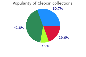
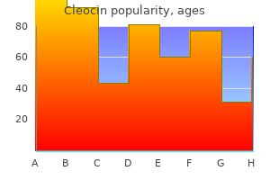
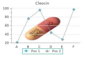
"Buy cheap cleocin line, skin care tips in urdu."By: Noreen A Hynes, M.D., M.P.H. - Director, Geographic Medicine Center of the Division of Infectious Diseases
- Associate Professor of Medicine
 https://www.hopkinsmedicine.org/profiles/results/directory/profile/0010761/noreen-hynes
Best cleocin 150mgThe straight sinus, sinus confluence, and tentorial apex are elevated above the lambdoid suture ("lambdoid-torcular inversion"). The transverse sinuses descend at a steep angle from the torcular herophili toward the sigmoid sinuses (36-36). The occipital bone could seem scalloped, focally thinned, and transformed with all posterior fossa cysts. If callosal dysgenesis is current, the lateral ventricles are extensively separated and may have unusually outstanding occipital horns (colpocephaly). The vermis is regular in size and morphology but is elevated and superiorly rotated with elevated tegmentovermian angle (18-45�). Retrocerebellar arachnoid cysts exert mass effect on the cerebellar hemispheres, which in any other case appear regular in morphology. They often appear related and without information of cyst wall histopathology may be tough to distinguish from each other. The finest approach is to describe the cyst by location, assess vermian growth together with measurement of the tegmento-vermian angle and the fastigium-declive line, and determine the mass effects-if any-on surrounding structures. Congenital Malformations of the Skull and Brain 1190 (36-41) Sagittal ultrasound in a new child infant exhibits a cystic posterior fossa fluid assortment. The tegmento-vermian angle is elevated, and the vermis appears rotated superiorly. The vermis is undamaged but rotated superiorly, and the tegmentovermian angle is elevated. Posterior Fossa Malformations 1191 (36-44) Coronal graphic of rhombencephalosynapsis reveals that no vermis is current in the midline of the cerebellum. Instead, the folia, interfoliate sulci, and cerebellar white matter are continuous across the cerebellar midline. Note absent vermis, transversely oriented folia, continuity of cerebellar white matter throughout the midline. Coronal and axial photographs present transverse folia and continuity of the cerebellar white matter across the midline (36-45). Images via the rostral fourth ventricle could show a diamond or pointed shape. Other forebrain anomalies embrace absent olfactory bulbs and corpus callosum dysgenesis. We now talk about a number of of those by which the abnormalities are largely defined by imaging features: rhombencephalosynapsis, Joubert syndrome, cerebellar hypoplasias, and unclassified dysplasias. Rhombencephalosynapsis is a midline mind malformation characterised by (1) a "lacking" cerebellar vermis and (2) obvious fusion of the cerebellar hemispheres (36-44). Severity ranges from delicate (partial absence of the nodulus and anterior and posterior vermis) to complete (the complete vermis, including the nodulus, is absent). Molar tooth problems are, a minimum of partially, "ciliopathies" with mutations of ciliary/centrosomal proteins that affect cell migration. The basic scientific presentation is a toddler with developmental delay, ataxia, and oculomotor and respiratory abnormalities. The fourth ventricle seems deformed with a thin upwardly (36-46) Axial graphic exhibits Joubert malformation. Thickened superior cerebellar peduncles round an elongated 4th ventricle form the traditional "molar tooth" signal. Posterior Fossa Malformations convex roof and lack of the normal pointed fastigium (3648A). Axial scans show the basic "molar tooth" appearance with foreshortened midbrain, slim isthmus, deep interpeduncular fossa, and thickened superior cerebellar peduncles surrounding an rectangular or diamond-shaped fourth ventricle. The superior vermis is clefted, and the cisterna magna may appear enlarged (36-48B). In rhombencephalosynapsis, the cerebellar hemispheres and dentate nuclei are fused across the midline, not cut up. A variety of findings together with enlarged, vertically oriented fissures or clefts (36-50), disordered or primitive foliation, lack of regular white matter arborization, gray matter heterotopias, and small cyst-like cavities in the subcortical white matter are a number of the many abnormalities seen in such circumstances (36-51). Selected References Chiari Malformations Chiari 1 Abu-Arafeh I et al: Headache, Chiari malformation type 1 and therapy options. Anomalies of the cerebral commissures are the most common of all congenital brain malformations, and corpus callosum dysgenesis is the single most common malformation that accompanies different developmental mind anomalies. Although they have an effect on very completely different parts of the forebrain, commissural and cortical malformations share an important function: they come up when migrating precursor cells fail to reach their target destinations. We start this chapter with a quick consideration of normal development and anatomy of the cerebral commissures, then focus on callosal dysgenesis as the most important anomaly that affects these white matter tracts. Their imaging detection, localization, and characterization have become more and more necessary in affected person management. Normal Development and Anatomy of the Cerebral Commissures Normal Development Normal Gross and Imaging Anatomy Commissural Anomalies Callosal Dysgenesis Spectrum Associated Anomalies and Syndromes Thick Corpus Callosum Malformations of Cortical Development Overview Three Stages of Malformations Malformations With Abnormal Cell Numbers/Types Microcephalies Focal Cortical Dysplasias Hemimegalencephaly Abnormalities of Neuronal Migration Heterotopias Lissencephaly Spectrum Cobblestone Lissencephaly Malformations Secondary to Abnormal Postmigrational Development Polymicrogyria Schizencephaly 1195 1195 1196 1197 1197 1200 1201 1201 1201 1202 1202 1204 1207 1210 1210 1212 1215 Normal Development and Anatomy of the Cerebral Commissures In this part, we briefly review normal growth of the commissures after which delineate their gross and imaging anatomy. Coordinated switch of knowledge between the cerebral hemispheres is important for normal mind function and happens by way of these three axonal commissures. Commissural improvement is a rigorously choreographed process in which axons from cortical neurons are actively guided across the midline to attain their targets within the contralateral hemisphere. Fiber bundles within the anterior and posterior callosum ultimately unite to type a single continuous structure, the definitive corpus callosum. The rostrum is the smallest section and connects the orbital surfaces of the frontal lobes. A outstanding anterior "knee"-the genu-connects the lateral and medial frontal lobes (37-1). White matter fibers curve anterolaterally from the genu into the frontal lobes because the forceps minor. Its fibers move laterally and intersect with projection fibers of the corona radiata. The isthmus is a shorter, barely narrower area that lies between the posterior body and splenium. The isthmus connects the pre- and postcentral gyri and auditory cortex with their counterparts in the contralateral hemisphere. Most of its fibers curve posterolaterally into the occipital lobes (37-2) as the forceps main. From the midline, it curves laterally within the basal forebrain and splits into two fascicles. Commissural and Cortical Maldevelopment the smaller more anterior bundle programs toward the orbitofrontal cortex and olfactory tract. It is a transversely oriented fiber bundle that crosses the midline within the posterior pineal lamina.

Buy 150mg cleocin with amexExercise regimens such as the Schroth methodology can be used alone or in combination with bracing to enhance the spinal deformity by growing muscle power and selling trunk elongation. This remedy is designed only for flexible curves and is restricted by the necessity for patient participation. Earlier surgical procedure is indicated if the patient has progressive respiratory or neurologic symptoms. Distraction maneuvers recontour the backbone utilizing short-term rods positioned along the spinal axis by screw or hook constructs anchored to the spine, ribs, and/or pelvis. Over time, the patient undergoes a collection of smaller surgical procedures to lengthen the rod and thus promote improved spinal alignment. Thoracic expansion is reserved for sufferers with a main deformity involving the thoracic cage, such as rib fusions or thoracic insufficiency syndrome. Compression-based methods use compressive implants to halt progress on the convex side of the curve. Staples or tethers are positioned throughout the expansion plates, and concave growth proceeds with the goal of curve balance. In follow, spinal development after a compression-based procedure can be unpredictable. In contrast to a posterior method, using an anterior compression-based method may get rid of the requirement for definitive fusion when a child reaches skeletal maturity. At mounted time intervals, this distraction-based construct is surgically lengthened by removing a securing clip, distracting the rods, and reapplying the clips to maintain the model new length (arrows). At fastened time intervals, this distraction-based assemble is lengthened by inserting an external control gadget on the pores and skin over the actuator (arrows). Magnetic attraction holds the two components in proximity, and the external control system elongates the actuator by a predetermined length without additional surgical procedure. Definitive surgical correction often combines spinal fusion with methods meant to enhance spinal alignment and stability. Obtaining axial aircraft correction usually requires osteotomies because the backbone has turn out to be stiff from the relative immobilization induced by the preliminary implants. Fusion surgical procedure seeks to halt the progress of the deformity by locking the spine into its present shape. Before fusion, spinal alignment could be improved in a quantity of planes by compression, distraction, and/or derotation of spinal implants mixed with Smith-Petersen or pedicle subtraction spinal osteotomies. These methods can be especially efficient when used asymmetrically or together. The possible long-term consequences of definitive fusion earlier than skeletal maturity include the crankshaft phenomenon, in which postfusion posterior development inhibition mixed with continued anterior progress causes a new or worsening deformity. Although delaying definitive surgical correction until the patient reaches skeletal maturity at all times is preferred, earlier surgical intervention could additionally be necessary. The affected person could require complicated spinal reconstruction involving irregular skeletal parts in isolation, as in a hemivertebrectomy, or in combination, as in vertebral column resection. Resection ideally is undertaken earlier than a secondary compensatory curve develops in order that the potential fusion length can be limited. The contraindications to resection embody evidence of spinal dysraphism at the similar degree, lack of ability to use internal or external bracing after resection, the presence of inflexible deformities above or beneath the extent, and vascular anomalies that will not be correctible. This elimination of the abnormal parts will shorten the backbone and is thus reserved for essentially the most severe spinal deformities. Complex reconstruction surgeries have a greater threat of bleeding and neurologic harm when compared with different spinal fusion procedures. It is recommended that these procedures be carried out by experienced surgeons with acceptable ancillary providers. Spinal progress happens in a guided style as the proximal and distal screws transfer along the tracks created by the rods (arrows). In this compression-based surgical intervention, the staples tether the spinal column alongside the curve convexity whereas allowing concave growth to stability the deformity. Unpredictable spinal growth and long-term sturdiness have restricted using staple correction in deformity correction. Patients with a neuromuscular or syndromic deformity typically undergo surgical correction at a comparatively early age because their disease is more doubtless to rapidly progress. These sufferers also have the best danger of postoperative morbidity because of associated medical circumstances. Complete removing of the hemivertebra adopted by a short-segment fusion permits curve correction and progress preservation in the remaining spinal column. Concomitant pulmonary pathophysiology necessitates the treatment of the spinal deformity with simultaneous preservation of the chest wall and lung growth. Unless the curve regresses or development ceases, preliminary remedy is designed to preserve bony progress until skeletal maturity, when definitive correction can happen. Concomitant pulmonary comorbidities often contribute to the general pathologic condition. Each etiology has distinctive pathologic traits and scientific findings that dictate the potential progression of the illness and administration choices. The objectives of remedy are to correct and/or control the deformity and protect skeletal progress potential till the affected person reaches skeletal maturity, at which time definitive correction can be undertaken, if essential. Distraction-based, guided-growth, or compression-based growth-friendly surgical methods can be used before definitive correction. The outcomes of intervention are troublesome to evaluate among teams of sufferers due to the heterogeneity of the pathologies. This study of human alveolar growth reported exponential growth within the first 2 years of life adopted by continued progress at a reduced fee in adolescence. This observational research of 202 Caucasian patients revealed diastematomyelia as the commonest intraspinal anomaly associated with congenital scoliosis. Gupta N, Rajasekaran S, Balamurali G, Shetty A: Vertebral and intraspinal anomalies in Indian population with congenital scoliosis: A research of 119 consecutive patients. This study of an Indian inhabitants reported that tethered cord was the most common intraspinal anomaly related to congenital scoliosis. Grauers A, Danielsson A, Karlsson M, Ohlin A, Gerdhem P: Family history and its affiliation to curve measurement and treatment in 1,463 patients with idiopathic scoliosis. Based on the self-assessment questionnaire responses of 1,463 sufferers with idiopathic scoliosis, the authors found larger curve sizes in patients with a optimistic family history of scoliosis, however no relationship between household history and intercourse or age of onset. A pattern toward improved deformity correction in sufferers with idiopathic pathology was found, along with normal longitudinal thoracic growth in all sufferers. The authors evaluate growth-friendly surgical treatment choices for youngsters with scoliosis, including distraction-based, guided-growth, and compression-based strategies. These remedies are geared toward curve control and upkeep of spinal and thorax development. Growth-friendly backbone implant techniques are categorized into three categories (distractionbased, compression-based, and guided-growth) to improve communication and facilitate comparative studies. This in vivo randomized research of magnetically managed growing rod expertise within a porcine mannequin indicated attainment of 80% of predicted spinal peak using distant distraction and an accelerated increase in spinal top after implant removal. This study of sufferers treated with Shilla progress steerage surgery reported a comparatively excessive complication rate however the capacity to achieve cheap curve control until definitive surgical correction. Vertebral physique stapling as an effective therapy in skeletally immature patients with idiopathic scoliosis was demonstrated in this study from a single institution.
Syndromes - Blood tests, including tests to check liver function and for viruses that might affect the liver
- Do NOT thaw out a frostbitten area if it cannot be kept thawed. Refreezing may make tissue damage even worse.
- Eat high-fiber foods
- National Hemophilia Foundation (NHF) - www.hemophilia.org
- Fever and diarrhea last for more than 3 days
- Amount swallowed
- Triple X Kit (also contains petroleum distillates)
Buy cheap cleocin lineThis abnormality normally occurs spontaneously in the course of the formation of reproductive cells (eggs and sperm) within the parents. Diagnosis is normally achieved via a history, physical examination, serum hormone ranges, and genetic testing (either before or after birth). Identification of this condition is often delayed until late childhood or early adolescence if the clinical presentation is extra subtle, but chromosomal evaluation can affirm the diagnosis. Early remedy permits for early hormone alternative to reduce issues and detect complications. Diagnostic procedures consist of a historical past, physical examination, hormone ranges, and chromosomal testing. Treatment consists of male hormone substitute to promote secondary intercourse characteristics. Psychological counseling and help could additionally be helpful to the patient and the parents. The syndrome often becomes apparent at puberty when testicles fail to mature, rendering affected boys infertile. These challenges embrace hypoxia, nutritional changes, an infection, irritation, and chemical substances. Cells adapt to the challenges in an attempt to prevent or limit injury as well as dying. Benign tumors are less more probably to trigger problems within the host or metastasize besides in phrases of location. Genetic and congenital disorders can develop from elements that disrupt regular fetal improvement or work together with defective genes. Genetic and congenital issues could additionally be present at start or may not seem till later in life. Exploring these basic cellular and genetic ideas and points lays the foundation for understanding where disease begins. Diagnosis and misdiagnosis of necrotizing soft tissue infections: Three case reviews. Humans can arm themselves with an arsenal of well being behaviors that may help defend towards these adversaries, but people usually enhance their vulnerability to hurt through different behaviors. All patients we encounter as healthcare suppliers are affected by this fixed state of warfare. Healthcare suppliers can establish these individuals in danger or beneath attack and assist them take up arms to defeat these persistent adversaries. Stress can arise from many occasions, even these that might be perceived as positive. Understanding the nature of stress and the consequences that it may possibly have on the physique is vital for healthcare providers in their interactions with sufferers. The Stress Response Hans Selye first described the bodily modifications related to stress within the Thirties. Selye described this protective stress response as the general adaptation syndrome, which is a cluster of systemic manifestations that symbolize an try to deal with a stressor. Several components can have an result on adaptation, together with pure reserves, time, genetics, age, gender, well being standing, vitamin, sleep�wake cycles, hardiness, and psychosocial components. The alarm stage consists of the generalized stimulation of the sympathetic nervous system resulting in the launch of catecholamines and cortisol, also recognized as the fight-or-flight response. Cortisol ranges and the sympathetic nervous system return to regular, causing the fightor-flight signs to disappear. The body either adapts or alters its workings in an try and restrict issues or turn out to be desensitized to the stressor. If the stressor is extended or overwhelms the body, the exhaustion phase is initiated. During this part, the physique turns into depleted and damage may seem, as homeostasis can now not be maintained by way of compensatory mechanisms. The local adaptation syndrome is the localized version of the final adaptation syndrome. In this syndrome, the physique makes an attempt to limit the harm associated with the stressor by confining the stressor to one location. An instance of this response may be seen within the native inflammatory reaction that outcomes from tissue trauma. Although the stress response is predictable to some extent, particular person variability exists due to conditioning elements. These conditioning components could embrace genetics, age, gender, life experiences, dietary standing, and social assist. The constructive presence of those factors can restrict or remove the chance of harm, disease, or dying. The implementation of a number of coping methods also can decrease and remove negative stress effects. These methods embody way of life modifications such as physical exercise, sufficient sleep, and optimal dietary standing. Unfortunately, maladaptive coping strategies might generally be used as an alternative of such optimistic methods. These maladaptive strategies trigger more issues than advantages and embrace actions corresponding to smoking, substance abuse, and overeating. Healthcare professionals can help patients to replace these unfavorable methods with extra positive ones. The immune system is answerable for protecting the physique towards an array of microorganisms. Fundamental to a properly functioning immune system is the flexibility to acknowledge and respond to a international agent, or antigen. Innate and Adaptive Defenses the immune system takes multiple approaches to protect the physique from antigens, including the utilization of innate and adaptive defenses. Innate immunity provides quick safety and is nonspecific, meaning it provides safety towards all invaders. Connective tissue containing many lymphocytes; transports immune cells, antigen-presenting cells, fatty acids, and fats; filters body fluids. Infection-fighting brokers; often the first to arrive on the scene of an an infection; attracted by varied chemicals launched by infected tissue; escape from the capillary wall, migrate to the location of an infection, and phagocytize microorganisms. White blood cells that bind immunoglobulin E (IgE) and release histamine in anaphylaxis. White blood cells that replenish macrophages and dendritic cells in regular states and reply to inflammation by migrating to infected tissue to become macrophages and dendritic cells; their conversion elicits an immune response. White blood cells within tissues, produced by differentiation of monocytes; phagocytize and stimulate lymphocytes and other immune cells to respond to pathogens.
Effective 150 mg cleocinWhen considering sensory perform, C5 supplies the higher outer arm, C6 the thumb, C7 the lengthy finger, C8 the little finger, T1 the medial forearm, T10 the periumbilical area, L1 the groin area, L2 the anterior thigh, L3 the knee, L4 the medial malleolus, L5 the nice toe, S1 the small toe, S2 the posterior thigh, and S3-S5 the perianal region. Both the proximal and distal radicular arteries anastomose at the proximal one-third of the spinal nerve root throughout the foramen, resulting in a area of potential vascular insufficiency. The innermost pia mater permits exchange of metabolites within the cerebrospinal fluid. The mechanical compression of a nerve root can end result in vascular compression, which can lead to the development of the traditional symptoms of radiculopathy. The cauda equina is an organized structure containing the lumbar and sacral nerve roots, and a quantity of other of the spinal nerve roots are organized into plexus constructions. The cervical plexus is composed of the ventral rami of C1C4; the brachial plexus is formed by the anterior rami of C5-T1; and the sacral plexus is made up of the lumbosacral trunk (L4, L5) and the S1, S2, S3, and S4 anterior rami. The preganglionic neurons of the sympathetic system are situated between C8 and L4. The sympathetic facilities control the cardiovascular and bronchopulmonary methods, sweat gland perform, vasomotor exercise, anorectal and bladder continence, and ejaculation. Horner syndrome is a clinical condition that can outcome from injury to the cervical or first thoracic sympathetic chain, which runs along the lateral borders of the vertebral physique. Injury can lead to a mixture of indicators corresponding to drooping of the upper eyelid (ptosis), contraction of the pupil (miosis), retraction of the attention (enophthalmos), and absence of sweating (anhidrosis). Hypogastric plexus injury (often where it lies anterior to the L1-S5 disk) can lead to urogenital issues similar to retrograde ejaculation. Autonomic dysreflexia results from spinal cord harm above the level of the sympathetic splanchnic outflow (T6) and leads to headache, sweating, flushing, and hypertension. The parasympathetic nervous system controls a big selection of visceral capabilities, including peristalsis and bladder wall contraction. The parasympathetic system also controls rest of sure easy muscular tissues such as those that regulate arterial blood move and penile erection. Many visceral parasympathetic features are carried through the vagus nerve and thus perform unopposed by sympathetic outflow in cases of spinal wire harm. Disruption of the sacral signaling pathways can impair crucial autonomic features similar to bladder and defecation management and sexual arousal. They subsequently enter an ascending fiber tract, such as the dorsal column pathways, which include the fasciculus gracilis and fasciculus cuneatus. The fasciculus gracilis controls the lower trunk and lower limbs, whereas the fasciculus cuneatus controls the higher trunk and upper limbs. The dorsal pathways management a variety of discriminative sensory functions, together with two-point discrimination, detection of speed, path of motion, and evaluation of cutaneous pressure. Its axons decussate to the ventrolateral column and terminate within the ventral posterolateral and central lateral nuclei of the thalamus. It originates within the motor cortex, with its axons forming the pyramidal tract, and most of its fibers decussate within the lower medulla to form the lateral corticospinal tract. The remaining fibers stay within the ventral funiculus and subsequently decussate in the ventral commissure. The corticospinal tract exerts refined motor management by way of its affect on different descending spinal pathways. It controls the sensory processing of pain, temperature, contact, and proprioception. The peripheral sensory receptors are specialised sense organs that connect with axons from the dorsal root ganglion. In this a half of the dorsal horn, the second-order neurons give rise to their processes, which carry signals to other areas of the brain and spinal wire. Somatic afferent fibers, along with the fibers controlling visceral sensation and ache, converge on the neurons of the substantia gelatinosa. Certain regions of the brain additionally supply substantial enter to impact neuromodulation within the substantia gelatinosa. This signaling pathway conveying the feeling of contact is also liable for controlling extra subtle sensory features similar to proprioception and two-point discrimination. The central department, making up the medial division of the dorsal root, splits after getting into the spinal cord. These monosynaptic connections have a excessive diploma of specificity, with muscle spindle afferents from a given muscle (in response to adjustments in muscle length and velocity) making contact solely with motor neurons that innervate the muscle of origin of the afferent fiber. The variability of reflex activity also is affected by temporal and spatial summation of excitatory inputs and inhibitory influences from different sources. Simple spinal reflexes demonstrate that neurons not solely are excited but in addition can be inhibited by sure inputs. In postsynaptic inhibition, the membrane potential of the postsynaptic neuron will increase, and the same excitatory enter is unsuccessful in depolarizing the neuron sufficiently to initiate an action potential. In presynaptic inhibition, a decreased quantity of excitatory transmitter is released from the presynaptic terminal. Spinal Cord Lesions When evaluating spinal wire lesions, you will want to classify the extent of the lesion. The time period tetraplegia generally refers to spinal wire harm on the cervical level, with a resultant neurologic deficit affecting the higher and lower limbs, trunk, and pelvic organs. The time period paraplegia refers to spinal twine harm at the thoracic, lumbar, or sacral ranges, with a resultant neurologic deficit affecting the lower limbs and pelvic organs; higher limb operate is preserved. In an incomplete injury, some motor or sensory perform below the harm stage is preserved. The Clarke nucleus is demonstrated in relation to the ventral and dorsal spinocerebellar tract and dorsal columns. Neurogenic shock leads to hypotension and relative bradycardia and could be fatal. It occurs as the outcomes of circulatory collapse from loss of sympathetic tone and is attributable to disruption of the autonomic pathway throughout the spinal twine. This leads to lack of sympathetic tone, decreased systemic vascular resistance, pooling of blood within the limbs, and hypotension. Spinal shock is a brief loss of spinal wire perform and reflex exercise below the extent of a spinal cord damage. It ends in a flaccid areflexic paralysis, bradycardia, hypotension, and an absent bulbocavernosus reflex. Spinal shock occurs because of hyperpolarization of neurons that remain unresponsive to mind stimuli. Central wire syndrome is the most typical and infrequently impacts aged people who maintain a minor extension harm. It is brought on by spinal wire compression and central wire edema and results in selective destruction of the white matter in the central area of the lateral corticospinal tract; the upper extremities are preferentially affected. Although central cord syndrome is related to a great prognosis, full functional restoration is rare. Anterior cord syndrome presents as motor dysfunction and dissociated sensory deficit below the extent of a spinal cord damage. Injury to the anterior spinal twine is usually brought on by direct osseous compression or injury to the anterior spinal artery.
Best 150mg cleocinThe pores and skin incision extends from the mastoid course of to the hyoid bone within the midline. Because the encountered neurovascular buildings are symmetric, the aspect of the method is dependent upon surgeon preference and pathology. At this stage of the process, it is very important establish three necessary neurovascular structures-the marginal mandibular nerve, deep to the parotid gland; the retromandibular vein, on the middle portion of the parotid; and the frequent facial vein, on the angle of the jaw. The submandibular gland is excised, and its duct is ligated to prevent fistula formation. The stylohyoid and the digastric muscular tissues are recognized and tagged after which indifferent from the hyoid bone, which helps in lateral retraction of the hyoid and the trachea/larynx. Care ought to be taken to keep away from harm to the hypoglossal nerve, which lies deep to the indifferent muscle tissue. The dissection is additional deepened in the plane between the carotid sheath laterally and the esophagus/larynx medially. The following seven important structures are recognized and ligated in a cranial to caudal direction: the facial artery and vein, the ascending pharyngeal artery and vein, the superior laryngeal artery, and the superior thyroid artery and vein. After performing the required procedures, the wound is closed over a drain and the digastric and stylohyoid tendons are repaired. Possible problems of the anterior retropharyngeal approach embrace damage to the esophagus, hypopharynx, or the neurovascular buildings beforehand mentioned. This strategy facilitates occiput to cervical and C1-C2 reconstruction procedures and is mostly indicated for situations corresponding to trauma, rheumatoid arthritis, infections, and tumors. Care should be taken to prevent fusion of the occipitocervical junction in a flexed or prolonged position as a end result of dysphagia, subaxial subluxation, and airway compromise can result. The palpable landmarks, including the occipital protuberance and the C2 and C7 spinous process, are marked, and the proper degree is confirmed under fluoroscopy. A midline incision is made extending from the occipital protuberance to the spinous strategy of C3. The superficial dissection is carried out strictly in the midline to reach the ligamentum nuchae. The rectus capitis and oblique capitis are subperiosteally elevated from the spinous course of and the lamina of C2, and a spotlight is then turned to the occipital bone the place subperiosteal dissection is carried out from the midline along the inferior nuchal line. Exposure is maintained with right-angle cerebellar or Gelpi retractors proximally and distally. The posterior tubercle of C1 is identified and subperiosteal publicity on both sides of the midline is carried out using curets. The vertebral artery runs alongside the cranial surface of the lateral third of the posterior arch. Meticulous surgical technique and avoidance of Bovie electrocautery and burring on the superior arch of C1 also are recommended. The exposure of the C1 lateral mass includes mobilizing the C2 dorsal nerve root caudally. Because this root, which lies on the junction of the posterior C1 arch and the lateral mass, is surrounded by an abundant perineural venous plexus, bleeding can make dissection in this space challenging. Without root transection, C2 neuralgia can happen after placement of C1 lateral mass screws in as many as 30% of cases. Alternatively, some surgeons use C1 pedicle screws with a starting point that lies on the posterior arch to avoid bleeding round and irritation of the greater occipital nerve that can outcome from lateral mass screw placement. Note the place of the greater occipital nerve because it pierces the C1-C2 membrane and programs laterally and superiorly. Exposure of the C1-C2 articulation for bone grafting generally requires retraction or sectioning of this nerve. Posterior Subaxial Cervical Approach in Laminoplasty and Laminectomy the posterior strategy to the subaxial cervical spine is often utilized in procedures similar to laminectomy, laminoplasty, and lateral mass fixation. For sufferers with substantial cord compression, a slight flexion alignment is initially most well-liked. If fusion is to be carried out, you will want to restore lordosis after decompression but before ultimate implant tightening. Surgical Steps the spinous processes of C2 and C7 are marked, and correct ranges are identified using fluoroscopy. The pores and skin and subcutaneous tissue can be infiltrated with a 1:500,000 epinephrine resolution to assist with hemostasis. The dissection is then deepened, maintaining strictly within the median raphe to keep away from bleeding and muscle injury. After the spinous course of is reached, further dissection is performed subperiosteally from distal to proximal utilizing electrocautery; self-retaining retractors are then placed bilaterally. Depending on the pathology handled, a laminectomy is performed in a piecemeal or en bloc trend. Specific Instructions: Laminoplasty When performing a laminoplasty, affected person positioning, skin incision, and lamina publicity are like those of the conventional posterior method, however care should be taken to protect the C2 and C7 attachments. Preservation of the C2 and C7 muscle attachments theoretically reduces lack of lordosis and neck pain brought on by mechanical instability after laminoplasty. Posterior Minimally Invasive Foraminotomy the minimally invasive strategy to the cervical backbone has been shown to scale back the length of hospital stays and postoperative pain treatment requirements. This strategy is mainly useful in treating radiculopathy secondary to lateral disk herniation with out instability and kyphotic deformity. A inclined position with a slight reverse Trendelenburg position can reduce bleeding. When small incisions are planned, fluoroscopy is relatively more necessary for identification and confirmation of the goal stage earlier than the incision is made. Surgical Steps A 3-cm midline skin incision is placed with the goal disk degree as the center. The superficial dissection is sustained by way of the midline until the cervical fascia is encountered, which is divided longitudinally within the midline to expose the ideas of the spinous course of. Further dissection is carried out subperiosteally elevating the paraspinal musculature from the lamina, spinous process, and aspect joint using a Cobb elevator and electrocautery. At this stage, a self-retaining or handheld retractor is placed to reflect paraspinal muscles from the target interlaminar area. A high-speed burr is used to take away the caudal edge of the higher lamina and the medial third of the side. This resection may be accomplished with a small Kerrison rongeur, however not extra than half of the side ought to be resected. Microscope magnification and safety of the nerve root with a small Penfield retractor is typically recommended. Bleeding from the perineural venous plexus can usually be controlled with hemostatic agents and cottonoid packing. The shaded region of the lamina, side area, pedicle, and vertebral body may be completely resected if a tumor or infection is present. This strategy is indicated within the therapy of tumors, infections, backbone fractures, and backbone dislocations. Intraoperative fluoroscopy or plain radiography is used to find the supposed surgical degree.
Discount cleocin 150mg without prescriptionHeadaches, habits changes, and seizures have been reported with supratentorial lesions. Total surgical removing is the treatment aim however may be troublesome because of adhesion of the cyst membrane to critical neurovascular structures. Age distribution is bimodal, with a big peak within the third and fourth many years and a smaller peak in the first decade. Density and signal intensity vary according to protein Neoplasms, Cysts, and Tumor-Like Lesions 882 content of the cyst fluid. Signal depth of cyst contents varies extensively, depending on imaging sequence and protein content material. A few instances of gentle posterior rim enhancement on the cyst-brain interface have been reported. The heart of the mass is just slightly offmidline, a typical resolution for a posterior fossa neurenteric cyst. Nonneoplastic Cysts 883 (28-24) Sagittal graphic reveals a small cystic lesion within the pineal gland. These lesions, usually seen in patients with obscure complaints and no signs referable to the pineal area, may be troublesome to each radiologists and referring clinicians. Theories include persistent coalescing embryonic pineal cavities and glial degeneration with cavitation. An straightforward approach to remember the conventional midline anatomic constructions in the pineal region-from top to bottom-is "well-known V. The common appearance is that of a easy, gentle, tan-yellow pineal gland that incorporates a uni- or multilocular cyst (28-25). The inner layer is a sharply outlined zone of finely fibrillar glial tissue with Rosenthal fibers. The inner surface of a pineal cyst cavity is often hemosiderin stained as the end result of intralesional hemorrhage. The incidence amongst ladies ages 21-30 years is significantly larger than in another group. A "thunderclap" headache may mimic symptoms of aneurysmal subarachnoid hemorrhage. Serial follow-up of indeterminate cystic lesions of the pineal region exhibits, in most lesions, no vital change in dimension or character over time intervals from months to years. Approximately 1-2% are very hyperintense, which can point out intracystic hemorrhage. Thin-section high-resolution sagittal and axial T2 scans are especially helpful for detecting and characterizing lesions within the anatomically complex pineal area. Neoplasms, Cysts, and Tumor-Like Lesions 886 Differential Diagnosis the commonest differential prognosis is normal pineal gland. Normal pineal glands typically contain a quantity of small cysts and can have nodular, crescentic, or ring-like enhancement. Pineocytomas can remain steady for many years with out vital change on serial imaging. Once a cyst has been identified as mendacity within the mind itself, the differential diagnosis is proscribed. The most typical parenchymal cysts-prominent perivascular areas and hippocampal sulcus remnants-are anatomic variants. Cyst formation may relate to blood-brain-barrier deficiency with peritumoral extravasation of water, electrolytes, and plasma proteins from altered microvessels. They are often uneven, may cause mass impact, and have regularly been mistaken for multicystic brain tumors. The cyst wall usually consists of gliotic brain with reactive astrocytes and lymphocytes. Most are solitary, however sometimes multiple loculated fluid collections are trapped on the tumor-brain interface. The widespread appearance is one or more "pools" of trapped fluid surrounding an extraaxial tumor mass (28-30). Enhancement is minimal or absent and customarily related to reactive inflammatory adjustments in the cyst wall, not tumor. They are additionally common within the subcortical and deep white matter as nicely as the midbrain and dentate nuclei of the cerebellum. For example, in the event that they occur within the subcortical white matter, the overlying gyri are enlarged with concomitant compression of adjacent sulci (28-38). Differential Diagnosis the major differential analysis is chronic lacunar infarction. Infectious cysts (especially parenchymal neurocysticercosis cysts) are often small. These remnant cavities-hippocampal remnant cysts-are regular anatomic variants (28-40). Small blood vessels are sometimes also included as the hippocampal sulcus forms, folds, and fuses. They appear as a "string of beads" with a quantity of small spherical or ovoid cysts curving along the hippocampus between the dentate gyrus and subiculum, simply medial to the temporal horn of the lateral ventricle. Etiology At 15 fetal weeks, the hippocampus usually unfolds and surrounds an "open" shallow fissure-the hippocampal sulcus-along the medial surface of the temporal lobe. The walls of the hippocampal sulcus gradually fuse, and the sulcus is eventually obliterated. When they occur within the temporal lobe, enlarged perivascular areas are found in the subcortical white matter of the insula and anterior tip of the temporal lobe, not medial to the temporal horn of the lateral ventricle. They are benign fluid-containing cavities buried inside the cerebral white matter. Porencephalic cysts communicate with the ventricle and are lined by gliotic or spongiotic white matter. Arachnoid cysts are extraaxial, not intraaxial, and are lined with flattened arachnoid cells. Connatal cysts are cystic ependyma-lined areas adjacent to the superolateral margins of the physique and frontal horns of the lateral ventricles. They are relatively common and usually innocuous lesions caused by coarctation or coaptation of the partitions of the frontal horns. Etiology Porencephalic cysts are encephaloclastic lesions, the tip result of a damaging process. Pathology Porencephalic cysts range in measurement from a number of centimeters to cysts that contain just about a whole cerebral hemisphere. Porencephalic cysts are deep, uni- or bilateral, smooth-walled cavities or excavations within the brain parenchyma. They are sometimes "full-thickness" lesions, extending from the ventricle to the glia limitans of the cortex (28-45).
Hedge Bindweed (Greater Bindweed). Cleocin. - Dosing considerations for Greater Bindweed.
- What is Greater Bindweed?
- Are there any interactions with medications?
- Fever, urinary tract diseases, constipation, increasing bile production, and other conditions.
- Are there safety concerns?
- How does Greater Bindweed work?
Source: http://www.rxlist.com/script/main/art.asp?articlekey=96179
Buy cleocin cheapThis decline was measured in a registry-based examine that discovered the speed of completion when utilizing 4 to five questionnaires was lower than 50% of that using two to three questionnaires. The demand for patient-centered outcomes additionally has contributed to the patient-level interpretation of present questionnaires. Most research report medical outcomes as adjustments in mean scores on varied consequence measures, with comparisons between scores before and after treatment or comparisons of scores between teams. Among suppliers, consciousness is growing of the necessity to gather outcomes knowledge; nevertheless, making an attempt to decide which devices should be used may be considerably overwhelming given the massive variety of devices described within the literature. Although good tools are available, they may not be relevant to the breadth of the population with spine situations, which includes people of various ages, occupations, leisure pursuits, and life. There is also an increasing preference for assessments which might be extra centered on the patient. Given the variety of questionnaires obtainable for spine patients, all with their very own advantages and downsides, it can be tempting to use lots of them. One growing enchancment in outcome knowledge assortment is electronic capture, which has improved data high quality by reducing issues encountered with paper administration. The greater responsiveness of questionnaires also probably will enhance cost-effectiveness research. All of those efforts will produce more dependable evidence that can be used to information patient care. Key Study Points Data collection in sufferers with spine ache is a frequently evolving process. A need exists for larger standardization of spine surgical procedure end result assessments, particularly the format and instructions supplied to facilitate knowledge pooling from a number of sources and in addition for more meaningful metaanalyses and different comprehensive critiques. The use of electronically administered end result assessments will probably proceed to enhance information assortment, including the usage of registries to accumulate giant numbers of sufferers, all finishing the same assessments, for knowledge analyses. A diverse international group of people with an interest in backbone care outcomes collaborated to develop a instructed core set of consequence assessments to be utilized in spine analysis. This evaluate of motion-preserving technology found that cost-effectiveness can range considerably primarily based on which extensively used general well being assessment was used within the calculation. This report highlights the challenges in reporting and interpreting costeffectiveness literature and the way inconsistent the literature could be. H�gg O, Fritzell P, Nordwall A; Swedish Lumbar Spine Study Group: the scientific significance of modifications in consequence scores after remedy for persistent low back pain. This study describes multiple methods of contemplating prices and a few of the obstacles in performing optimum cost analyses. Davidson M: Rasch analysis of three variations of the Oswestry Disability Questionnaire. The 5-year outcomes of patients with lumbar stenosis handled with an interlaminar gadget to provide spinal stability with out fusion were evaluated. This study investigated cost-effectiveness by figuring out the minimal quantity of improvement needed on an consequence assessment for the therapy to be thought of costeffective. Rather than requiring that a selected change in outcome scores be achieved, this research proposed determining a minimal worth be achieved on an evaluation for an outcome to be categorized as successful. Murrey D, Janssen M, Delamarter R, et al: Results of the possible, randomized, controlled multicenter Food and Drug Administration investigational system exemption study of the ProDisc-C whole disc substitute versus anterior discectomy and fusion for the therapy of 1-level symptomatic cervical disc illness. This examine used an digital model of the traditional ache drawing to examine the restoration sample of assorted sensations after surgical decompression of lumbar nerve roots. This examine reported that satisfaction scores may be substantially affected by factors other than the quality of care acquired, similar to misery. Based on a satisfaction survey typically used in health care, higher satisfaction for patients present process spine surgical procedure was associated to lower ache scores and a sense that a care provider spent more time with them. Although demand is rising for the inclusion of patient satisfaction in consequence reporting, satisfaction was not related to commonly used consequence assessments, hospital readmission, or morbidity rates. This complete evaluation reported on the price of backbone issues within the United States and the disproportionally low amount of research funding spent on backbone problems compared with varied other situations. This refutes conventional thought that new methods are inherently dearer. This study demonstrates how general health instruments can be used to examine the cost-effectiveness of spine care within the context of different problems generally accepted as cost efficient. A good price of patient acceptance was discovered for utilizing a cellular tablet to acquire patientcompleted questionnaires. Frennered K, H�gg O, Wessberg P: Validity of a computer touch-screen questionnaire system in again patients. This research highlights the need for rigorously worded and rigorously implemented definitions for knowledge factors such as reoperation in medical research. The reoperation rates varied considerably among the many similar sufferers based on the definitions utilized. This research found that comparable information values had been collected utilizing both phone interview or having the patient full the form in the clinic. Morris S, Booth J, Hegarty J: Spine Tango registry knowledge collection in a conservative spinal service: A feasibility study. This examine reported that the greater the variety of evaluations administered, the larger the speed of data loss. Those who take care of athletes should be familiar with harm prevention methods together with the on-field management of backbone accidents and return-to-play issues. The capability of an athlete to return to play depends on the pathoanatomy of the injury and the results of therapies. The danger for additional harm is the first consideration in the willpower if an athlete can return to sports activities participation. Keywords: athletic injuries; cervical fractures; return-to-play tips; group physicians Dr. Harris nor any instant family member has acquired something of value from or has stock or stock options held in a industrial company or establishment associated directly or indirectly to the topic of this chapter. Introduction Sports-related accidents are a typical explanation for unintended injury, accounting for approximately 5. Such fears have to be weighed towards the benefits that sports and leisure actions provide, nevertheless. Participation in sports activities is necessary for physical well being and overall well-being. Surgeons who present backbone care should perceive the mechanisms and patterns of injury in sport-specific settings, on- and off-the-field management of such injury, and the steps concerned in secure return to play. Sport-Specific Epidemiology Epidemiologic data on sports-related spine and spinal wire injuries varies by country. Spine injuries range from simple contusions and muscular strains to fractures with full spinal wire damage.

Buy generic cleocin 150mg lineIt is helpful to be familiar with the important rules of early care of spine trauma together with latest developments within this field. Keywords: preliminary resuscitation; neurologic evaluation; backbone imaging; backbone immobilization; traumatic spinal wire harm Dr. Kwon or a direct family member serves as a paid marketing consultant to Acorda Therapeutics. Bourassa-Moreau nor any quick member of the family has obtained anything of value from or has stock or stock options held in a commercial company or establishment associated directly or indirectly to the subject of this chapter. Introduction Early recognition of backbone trauma is important for the initiation of applicable care that protects the spinal cord from additional damage. Physicians caring for such patients early after their damage could also be answerable for immobilizing the affected person appropriately, analyzing and documenting the diploma of neurologic impairment, obtaining the necessary imaging studies, characterizing the nature of the backbone injury, and organizing specialised care if necessary. This article evaluations the essential ideas of early administration of backbone trauma, with an emphasis on incorporating recent knowledge within the area. Airway Control and Cervical Spine Protection the possibility that a trauma patient has sustained an unstable cervical backbone injury mandates cervical backbone immobilization and protection throughout early airway administration. If intubation is critical, it should be performed with the backbone maintained in impartial alignment and with as little movement as possible. The trauma staff should anticipate situations in which intubation with cervical spine immobilization may be challenging and make sure that acceptable experience for securing the airway (such as anesthesiology) is on the market. With accidents at C3 or larger, patients might expertise acute respiratory arrest on the damage scene, which requires pressing intubation and ventilation. Circulation Hypotension in the affected person with acute trauma is assumed to have a hemorrhagic etiology until proved otherwise. Irrespective of etiology, hypotension have to be aggressively corrected to minimize secondary ischemic injury to the spinal twine and other sensitive organs. The presence of voluntary movement within the four extremities should be established after full exposure of the affected person. The presence of tenderness, swelling, or a step deformity can suggest the presence of a posterior ligamentous damage and warrants advanced imaging. Perianal and anal sensation, anal tone, and voluntary contraction, as well as the bulbocavernosus reflex, should all be documented. An absent bulbocavernosus reflex means that spinal shock remains to be current, and the true extent of the neurologic impairment may not be totally appreciated. Characterizing the true extent of neurologic impairment within the early stages after damage is regularly sophisticated by concomitant injuries (including mind injury), pharmacologic sedation, or intoxication. Motor operate is assessed by measuring power in five key muscle groups within the upper extremities and five key groups in the decrease extremities. Sensory evaluation of light contact and pinprick sensation is accomplished all through 28 dermatomes. The interpretation of the completeness of injury relies on the presence of perianal and anal sensation, anal tone and voluntary contraction, and the bulbocavernosus reflex. The time required is challenging in the setting of acute trauma for surgeons whose time is limited, and patient participation is frequently inconceivable because of concomitant injuries, intoxication and/or sedation, or even language limitations. Imaging Assessment for Spine Trauma In the trauma setting, the primary question is whether imaging is actually required. C, Thoracic midsagittal T2-weighted magnetic resonance picture reveals T3-T4 disk material abutting against the spinal cord. D, Cervical midsagittal quick tau inversion restoration magnetic resonance image reveals multilevel cervical spinal stenosis. Cervical Spine Clearance Arguably, no other matter has raised as a lot concern and anxiety within the setting of spinal trauma than the means to clear the cervical backbone within the obtunded, unexaminable affected person, and specifically, what imaging studies are required to achieve reasonable assurance for cervical collar elimination from such sufferers. The patient reported steadily worsening pain and saddle paresthesia over the following 2 weeks. C, Midsagittal T2-weighted magnetic resonance picture reveals local compression of the conus medullaris. D, Lateral lumbar radiograph obtained following posterior decompression and lengthy (T9L5) instrumented fusion. Spine Ankylosis the extensive literature regarding imaging and the evaluation of stability is based on displacement and ligamentous integrity. Fractures of any diploma within the ankylosed backbone may be potentially extremely unstable because of the lengthy lever arms being utilized to the fracture site. In instances of ankylosing spondylitis or diffuse idiopathic skeletal hyperostosis, low-velocity injury (such as a fall from a standing height) can result in fractures that will seem trivial but are doubtlessly highly unstable. This degree of vigilance is crucial in the evaluation of these patients because preliminary imaging studies may not reveal the fracture, which, by advantage of the ankylosis, must fully disrupt both the anterior and posterior columns, as with a long bone fracture. In sufferers with backbone ankyloses, the entire spine must be absolutely imaged as a outcome of concomitant spine fractures are widespread and the historical past and bodily examination are sometimes unreliable. Individualized immobilization utilizing sandbags, beanbags, towels, and straps is safer than utilizing standard collars and braces, which can further lengthen and displace a fracture initially brought on by hyperextension. Patients with ankylosing spondylitis should be carefully monitored due to the excessive risk of epidural hematoma and related delayed neurologic damage. Anesthesiology consultation is suggested if any surgical procedure is deliberate due to the difficulty in securing the airway. Therapeutic Modalities in Spine Trauma After a spine injury has been identified, the surgeon should assess the diploma of instability to decide whether or not surgical fixation is required or exterior immobilization with some type of orthosis shall be sufficient. Clearly, not all backbone fractures require surgical intervention, and the decision making regarding surgical procedure typically considers the want to provide stability, the necessity to decompress neurologic parts, and affected person components that may affect the choice of surgical or nonsurgical remedy (such as medical comorbidities and physique habitus). Classification techniques such because the Subaxial Cervical Spine Injury Classification had been established to help guide the clinician on this decision-making course of by accounting for the degree of mechanical instability and the extent of neurologic impairment. Bracing and External Immobilization Bracing remains a typical definitive treatment of many spine injuries deemed sufficiently stable to heal with out surgical fixation. A 2016 systematic review reported on the effectiveness of cervical orthoses for immobilizing the backbone. By comparability, craniothoracic units limit 60% to 80% of cervical movement in all directions. Caution must be exercised for making use of halo vests within the aged inhabitants as a end result of these devices have been associated with vital complications, including aspiration, pneumonia, cardiorespiratory occasions, and death. Similarly, for an osteoporotic compression fracture, an exterior orthosis is a suitable remedy possibility, though robust medical evidence to help its use is lacking. Closed Reduction of Spine Fractures and Dislocations the objective of closed discount is to realign a dislocated backbone and probably relieve any ongoing spinal cord compression. When and the method to cut back a cervical side dislocation continues to be discussed due to the chance that a disk herniation might be displaced into the spinal cord. In a affected person with complete quadriplegia and ongoing cord compression, a fast closed reduction directly for imaging could present substantial reduction, particularly if performed very early after damage. At some establishments, it might be faster to perform an anterior cervical diskectomy and open discount than to arrange the necessary gear and assemble the required employees to conduct a closed discount in an awake affected person. The technique for closed reduction of a cervical aspect dislocation in an awake, cooperative particular person entails pharmacologic sedation, muscle rest, close monitoring of respiratory standing, traction, and repeated radiographic evaluation. Traction weights of 15 lb for the head and an additional 10 lb per degree above the dislocation have been beneficial, however secure discount with weights properly over a hundred lb even have been described.
Generic cleocin 150mg mastercardAlterations within the regular microenvironment of pituitary stem cells may trigger uncoordinated proliferation and subsequent formation of pituitary adenomas. Adenomagenesis is a multistep, multicausal course of that features both initiation and development phases. A number of activated oncogenes and loss of tumor suppressor gene features are concerned. In addition, several endocrine components at either the hypothalamic or systemic level may induce adenohypophysial cell proliferation. Carney complicated is related to spotty pores and skin pigmentation, myxomas, endocrine tumors, and schwannomas. Tumors or nodular hyperplasia of a number of endocrine glands result in hypersecretory syndromes such as acromegaly, hyperprolactinemia, and Cushing syndrome. They sometimes develop development hormone-secreting adenomas and prolactinomas, often in childhood. The second, much larger group has adult-onset illness and extra diversified forms of adenoma. Adenomas differ in dimension from microscopic lesions (25-50A) (25-50B) to large tumors more than 5 cm that invade the skull base and prolong into a number of cranial fossae. Macroadenomas are red-brown, lobulated lots that usually bulge upward via the opening of diaphragma sella (25-43) or, less commonly, extend laterally toward the cavernous sinus. Histologic examination exhibits a uniform inhabitants of round, polygonal, or elongated cells Pathology Location. Reported ectopic websites embody the sphenoid sinus (the most typical site), nasopharynx, third ventricle, and suprasellar cistern. Adenoma classification is now based on immunohistochemical profile and scientific presentation. Female patients with prolactinomas current with amenorrhea-galactorrhea syndrome, whereas male patients current with hypogonadism and impotence. Patients with corticotroph tumors present with Cushing illness or Nelson syndrome (rapid enlargement of an adenoma following bilateral adrenalectomy). Although pituitary adenoma progress rates are quite variable, most enlarge slowly over a interval of years. Treatment options are numerous and include surgical resection, medical management, stereotactic radiosurgery, and standard radiation remedy. Approximately 60% of patients present process surgery have macroadenomas, and 40% have microadenomas. Note, however, that "large" pituitary adenomas could erode and extensively invade the cranium base, mimicking metastasis or aggressive an infection. Macroadenomas are often isodense with grey matter, however cysts (15-20%) and hemorrhage (10%) are common. The posterior pituitary "shiny spot" is absent (20%) or displaced into the supradiaphragmatic cistern (80%) on T1-weighted sagittal scans. Fluid-fluid ranges may be present but are more common in patients with pituitary apoplexy. Unless they hemorrhage, small microadenomas may be inapparent on normal nonenhanced sequences. Others enhance extra strongly and will turn out to be isointense with the enhancing pituitary gland, rendering them nearly invisible. This discrepancy in enhancement timing can be exploited through the use of thin-section coronal dynamic contrastenhanced scans. Fast picture acquisition during contrast administration can typically discriminate between the slowly enhancing microadenoma and quickly enhancing regular gland. Differential Diagnosis the differential diagnosis of pituitary adenoma varies with dimension and patient demographics. The main differential diagnosis of pituitary macroadenoma is pituitary hyperplasia. The height of the gland is usually no much less than 10 Sellar Neoplasms and Tumor-Like Lesions mm except the patient is pregnant or lactating. Less generally, end-organ failure (such as hypothyroidism) ends in compensatory pituitary enlargement. As adenomas are very rare in children, if a prepubescent female affected person or young male patient has an "adenoma-looking" pituitary gland, endocrine work-up is mandatory! Tumors that may resemble pituitary adenoma embody meningioma, metastasis, and craniopharyngioma. Meningioma of the diaphragma sellae can usually be recognized as clearly separate from the pituitary gland under. Metastasis to the stalk and/or pituitary gland from an extracranial major neoplasm is uncommon. Most pituitary metastases are secondary to unfold from adjacent bone or the cavernous sinus, usually occurring as a late manifestation of known systemic tumor. Craniopharyngioma is the most common suprasellar tumor of childhood, whereas pituitary adenomas in children are uncommon. Often in adults with craniopharyngioma, the pituitary gland may be identified as anatomically separate from the mass. Because of this rarity, even probably the most aggressive-looking pituitary tumors are statistically much more likely to be adenomas than carcinomas. Nonneoplastic entities that can mimic macroadenoma include aneurysm and hypophysitis. An aneurysm arises eccentrically from the circle of Willis and is often not within the midline immediately above the sella. Hypophysitis is far much less frequent than macroadenoma but can seem nearly similar to an adenoma on imaging research. Lymphocytic hypophysitis-the commonest type-typically happens in peripartum or postpartum female sufferers or as an autoimmune hypophysitis in sufferers treated with immunomodulating therapies. Pituitary microadenoma could additionally be troublesome to distinguish from incidental nonneoplastic intrapituitary cysts such as Rathke cleft cyst or pars intermedia cyst. Microadenomas improve; cysts are seen as nonenhancing foci throughout the intensely enhancing pituitary gland. Conventional histologic standards for malignancy (necrosis, nuclear atypia, pleomorphism, mitotic activity) are insufficient for diagnosis. Only documentation of craniospinal metastases or systemic tumor unfold can verify the diagnosis. Pituitary Blastoma Pituitary blastoma is a just lately described pituitary tumor in neonates and infants characterised by large glandular constructions that resemble Rathke epithelium and adenohypophysial cells. Arrested pituitary development and unchecked proliferation are the probably etiology of this unusual tumor.
Discount cleocin 150mg onlineThis procedure can be carried out unilaterally or bilaterally, depending on surgical indications. At this point within the process, the paraspinal muscle tissue may be held laterally with self-retaining retractors. Exposure lateral to the aspect joints may be completed if pedicle screws are deliberate or entry to the intertransverse region is desired for bone graft placement. When using typical pedicle screws, the multifidus tendons could be launched from the lateral aspect capsule with electrocautery. This facilitates gentle retraction of the muscle tissue lying over the transverse processes, which could be elevated rather easily with a Cobb elevator. Care ought to be taken across the superior and inferior margins of the facet as a end result of arterial perforators could be a nuisance if not acknowledged and cauterized. For pedicle screw placement via the cortical bone trajectory, the publicity solely needs to attain the lateral aspect of the pars interarticularis. This spares the muscle attachments alongside the lateral side joint, which aids in achieving a minimally invasive midline dissection. After the specified procedure is completed, the muscle, fascia, subcutaneous tissue, and pores and skin are closed in particular person layers. Advantages and Limitations Advantages of the posterior midline method are surgeon familiarity, clear appreciation of the anatomy, and little or no threat of neurovascular harm. Limitations include attainable extreme blood loss, intensive soft-tissue injury, and extreme postoperative pain. Some authors report that L4-L5 can be approached by way of increased lateral flexion of the patient, but the threat of L4 nerve damage also needs to be thought of in preoperative planning and intraoperative execution. In addition, the diaphragmatic crus or diaphragm itself might inhibit entry on this area. Transdiaphragmatic access is possible; nonetheless, the surgeon must be ready to place a chest tube in this setting. The use of an adjustable table with a break positioned at the disk area of curiosity (especially at decrease lumbar ranges and thoracolumbar levels) may facilitate safe entry. Care must be taken to flex the ipsilateral hip, which can help in mobilization of the psoas with out putting unnecessary strain on the lumbar plexus during retractor placement. A 3-cm incision is placed on the left flank, and electrocautery is then used to reduce via the exterior oblique fascia. Blunt dissection with a finger or peanut is used to attain the retroperitoneum via the external oblique, internal indirect, and transverse abdominis muscles. Branches of the subcostal iliohypogastric and ilioinguinal nerves may be encountered either running freely within the retroperitoneum or, more commonly, between the interior indirect and transversus abdominus. To reach the psoas, dorsal to ventral blunt dissection is used to move the peritoneum away from the surgical airplane. Before passing through the psoas, the anterior vessels and posterior lumbar plexus are checked. To guarantee their security, the psoas must be separated between the center and anterior third of the muscle, and the belly contents along with peritoneum are protected by placing handheld retractors. After the psoas muscle is visualized, sequential tubular dilators are handed by way of the muscle to attain the disk house. A retroperitoneal strategy to the lumbar spine from L5-S1 to L1-L2 is achieved via a single skin incision. For multilevel procedures, an indirect skin incision of 5 to 10 cm is created consistent with the fibers of the exterior oblique muscle alongside the lateral wall of the stomach. Abdominal muscles can be bluntly separated with minimal cautery after dividing their fascia. The peritoneum is separated from underlying retroperitoneal buildings by blunt finger dissection, and the abdominal contents are retracted anteriorly. The psoas muscle and the genitofemoral nerve are visualized, and the focused disk space is then approached between the left psoas and the aorta. A spinal needle or Kirschner wire is positioned in the disk house to verify the surgical level fluoroscopically. Importantly, when approaching the L5-S1 degree, the disk is removed lateral to the widespread iliac vessels quite than within the bifurcation. Guidewires are inserted into the pedicles by way of 1- to 2-cm paramedian incisions, and cannulated pedicle screws are positioned over the guidewire. If substantial signs are current bilaterally, the contralateral facet can be immediately decompressed by depressing the thecal sac anteriorly and "crossing over" to the other side and performing the decompression. Sequential soft-tissue dilators are then docked on the intervening facet and expanded to a desired working diameter of roughly 24 to 28 mm. Various further retractors could be inserted over the tubular retractors to allow even higher visibility. The decompression of the lateral recess and foramen is performed via the ipsilateral side and pars interarticularis. Contralateral decompression of the spinal canal could be carried out by angling the retractor blades to the opposite aspect or by inserting the retractor on the contralateral side and repeating this step. Pedicle screws are placed in a percutaneous style on the contralateral side to complete the assemble. A, With the affected person within the lateral decubitus position, the skin is incised in diagonal style just proximal to the iliac crest. Below the subcutaneous fat, the external indirect muscle is encountered and could be divided parallel to the muscle fibers (dotted line). B, Below the exterior oblique muscle, the interior oblique muscle is encountered and could be divided parallel to the muscle fibers (dotted line). C, Below the interior indirect muscle, the transversalis fascia is encountered and could be divided consistent with the skin incision. D, the retroperitoneal contents are gently retracted anteriorly to expose the psoas muscle and anterolateral side of the lumbar backbone. Anterior Lumbar Interbody Fusion Anterior lumbar interbody fusion approaches the disk area from a nearly direct anterior retroperitoneal approach. Because this process provides extensive entry to the disk house, it can be used to treat an unlimited spectrum of conditions, including degenerative situations, deformities, spondylolisthesis, and failed posterior surgery (such as pseudarthrosis). To facilitate pure lumbar lordosis, a roll or a bump is placed underneath the lumbar spine. A vertical, paramedian skin incision may be needed for multiple levels; nonetheless, a low transverse Pfannenstiel incision can be used for exposure to L5-S1 and sometimes for L4-L5. After the pores and skin incision, additional dissection via the fatty layer is performed using electrocautery to reach the anterior layer of the rectus sheath, which is then incised vertically. The peritoneum is separated using blunt finger dissection to create the retroperitoneal aircraft. The peritoneum is retracted from the left aspect towards the middle using handheld retractors to attain the anterior floor of the great vessels. Dissection is carried out anterior to the psoas muscle; care have to be taken to protect the genitofemoral nerve, which lies on the anterior floor of the psoas. Each anatomic level has a different relationship to surrounding neurovascular structures.
References - Summit, R. L. (1993). Urogynecologic causes of chronic pelvic pain. In F. W. Ling (Ed.), Obstetrics and gynecology clinics of North America: Contemporary management of chronic pain (p. 685). Philadelphia: W. B. Saunders. Surrey, E., & Judd, H. (1992). Reduction in vasomotor symptoms and bone mineral density loss with combined norethindrone and long-acting gonadotropin-releasing hormone agonist therapy of symptomatic endometriosis: A prospective randomized trial. Journal of Clinical Endocrinology and Metabolism, 75, 558.
- Lumen N, Oosterlinck W, Decaestecker K, et al: Endoscopic incision of short (<3 cm) urethral strictures after phallic reconstruction, J Endourol 23(8):1329n1332, 2009.
- Kang HJ, Imperato-McGinley J, Zhu YS, et al: The first successful paternity through in vitro fertilization-intracytoplasmic sperm injection with a man homozygous for the 5?-reductase-2 gene mutation, Fertil Steril 95:2125, e5n8, 2011.
- Luo Y: A potential modality for interstitial cystitis: intravesical use of antiinflammatory peptide RDP58, J Urol 173(2):340, 2005.
|
|


