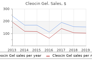
"Cleocin gel 20gm mastercard, skin care zamrudpur."By: J. Matthew Brennan, MD - Associate Professor of Medicine
- Member in the Duke Clinical Research Institute
 https://medicine.duke.edu/faculty/j-matthew-brennan-md
Order discount cleocin gelAs the remaining intestine undergoes adaptive changes, steady enteral feedings turn into needed. As this latter process may be pretty lengthy, direct gastric access through gastrostomy is fascinating. Other surgical pathology In any neonatal or infant situation during which a protracted ileus or partial luminal occlusion. Duodenal obstruction Congenital duodenal obstruction is normally related to proximal duodenal dilatation and atony in addition to gastric dilatation. Total parenteral nutrition and nasogastric decompression are generally efficient in postoperative administration. As said, nasogastric tubes tend to drain better than gastrostomies within the quick postoperative period. However, a gastrostomy can remove the need for long-term nasogastric or orogastric intubation and the problems related to placement and maintenance of these tubes. Gastrostomies intervene less with oral feedings than do nasogastric tubes, though the newer, smaller catheters are better tolerated. Gastrostomies are most popular over jejunostomies because the latter are less physiological and extra susceptible to mechanical problems. Gastrostomies, as properly as any other enteral tubes placed in infants, are related to a protracted list of potential early and late complications. Gastrojejunal tubes could eliminate the need for antireflux surgical procedure in patients with gastroesophageal reflux. Jejunostomies have been discovered to present more complications, such as inside hernia and volvulus (the latter notably within the Roux-en-Y jejunostomies or the "dissociation" approach bypassing the stomach as described by Bianchi). A notable morbidity after gastrojejunal tube placement has been described starting from persistent reflux to dislodgement and intestinal perforation. Normal feeding progressively returned, and some weeks after this picture was taken, the gastrostomy might be removed. The major indications in infants are swallowing difficulty secondary to central nervous system lesions as nicely as different abnormalities of deglutition, feeding supplementation, large-volume medications, and persistent malabsorption syndromes. Because the neurologically impaired kids incessantly have foregut dysmotility and gastroesophageal reflux in addition to swallowing difficulties, antireflux procedures are at occasions added to gastrostomies. There are conflicting outcomes about an increased danger of pathologic gastroesophageal reflux after gastrostomy tube placement in neurologically impaired children, leading to divergent suggestions of simultaneous antireflux surgical procedure. These techniques and their many variations are primarily based on three basic principles: Stamm gastrostomy 163 1. Formation of a serosa-lined channel from the anterior gastric wall round a catheter. This catheter is positioned in the abdomen and exits either parallel to the serosa, as within the Witzel approach, or vertically, as within the Stamm or Kader approaches. Construction of a tube or conduit from a full-thickness gastric wall flap, leading to the skin surface. A standard gastrostomy tube or a skin-level device may be placed beneath local anesthesia, although common anesthesia is most well-liked because abdominal wall leisure is required. After the tract is nicely healed, this stoma is suitable for the passage of dilators or guide wires. One of these is the percutaneous endoscopic "push" technique, which is performed with assistance from needledeployed gastric anchors or T-fasteners, and the Seldinger technique of guide-wire introduction followed by progressive tract dilatations. These are primarily expansions of the aforementioned strategies, considerably increasing the alternatives of gastric entry techniques out there to surgeons managing infants. When possible, a nasogastric tube is inserted for decompression and to help establish the abdomen, if necessary. This incision must be neither too excessive, as a end result of it will bring the catheter too near the costal margin, nor too low, avoiding the colon and the small bowel. However, this strategy is much less fascinating because the linea alba is the thinnest area of the stomach wall. Fascial layers are incised transversely and the rectus muscle retracted or transected. The website should be neither too excessive, because this would intervene with a fundoplication should one be wanted in the future, nor too low, as a outcome of stomas on the level of the antrum are prone to leakage and pyloric obstruction by the catheter. The surgeon must not place the catheter too near the higher curvature, to keep away from the so-called gastric pacemaker and to decrease the potential for gastrocolic fistula. The gastrotomy, at the heart of the purse string, is made sharply via the serosa and muscular wall of the abdomen. Other appropriate catheters are the Malecot or the T-tube, but both have the disadvantage of turning into more easily dislodged. We avoid the Foley or balloon-type catheters because the primary lumen is proportionately smaller and the balloon occupies extra intragastric area. Long balloon-type catheters, which can rupture, even have a larger propensity for distal migration into the small bowel. The continuous monofilament suture, used to anchor the abdomen to the anterior stomach wall, has been partially positioned. Although some surgeons convey the catheter out by means of the primary abdominal incision, wound complications which will happen in this setting tend to be more complex. The skin could be approximated with both interrupted or steady 5-0 or 6-0 subcuticular sutures. The catheter is firmly secured with two sutures of 3-0 or 4-0 artificial monofilament thread. These sutures are removed after 1 week, and a small crossbar is positioned loosely to forestall distal catheter migration. Conversion of a long tube to a button can be carried out after a agency adherence between gastric and belly wall is established. This offers a 360� fixation of the stomach to the anterior stomach wall with a watertight seal. The scope is inserted and advanced slowly into the abdomen, at which point the sunshine is seen by way of the left-upper-quadrant stomach wall. With the gastroscope in place, insufflation distends the stomach, apposes it against the anterior belly wall, and displaces the colon downward. When the room lights are dimmed, the gastric contour is clearly seen, particularly in babies. The subcuticular closure, adhesive strips, and secured gastrostomy catheter are depicted. The immobilizing sutures are eliminated after several days, and a small crossbar is placed to stop distal catheter migration. An alternative is the placement of a "button" or a balloon-type skin-level gadget, as an alternative of an extended tube, at the initial process. Gentle traction on the catheter assures that its intragastric place is maintained. The posterior rectus sheath is closed with a working suture of 4-0 absorbable, synthetic materials.
Syndromes - Both vaccines protect against the two types of HPV that cause most cases of cervical cancer. Other, less common types of HPV can still cause cervical cancer.
- Lower end of the thigh bone. This bone is called the femur. The replacement part is usually made of metal.
- Defects in your heart valve are causing major heart symptoms, such as chest pain (angina), shortness of breath, fainting spells (syncope), or heart failure.
- Does anything help? Like heat or massage to the lower abdomen?
- Echo-Doppler
- Examine the scalp and hair for moving lice and eggs (nits).
- High altitudes
Cleocin gel 20gm cheapIf through the initial exploration the nerve is famous to be transected and restore deferred, then the nerve endings should be identified and tacked right down to the adjacent tissue; doing so reduces the extent of retraction and makes it simpler to establish the ending later. Achieving a clean, d�brided wound with sufficient circulation and correct skin closure takes priority over performing the restore as an acute procedure. Neurorrhaphy should be undertaken solely when the medical situation and the environmental milieu of the nerve are as close to optimum as potential. Nerves by no means heal, a lot much less regenerate, in a grimy, contaminated, poorly vascularized wound. Such accidents range from blunt trauma to direct through-and-through penetration of the nerve. Depending on the extent of the harm and the quantity of scar shaped, important functional nerve loss can occur. Other essential elements that determine the diploma of harm are the anatomical degree of injection inside the nerve and the neurotoxicity of the drug. At acute exploration, exposure of the sciatic nerve confirmed it to be acutely swollen and discolored, instantly in the trajectory of the bullet. A variety of medical research have examined the toxicity of injectable drugs; for extra detailed info, the reader is referred to the report by Gentili and Hudson. The main issue that can decide the amount of injury is once again the sort of drug injected. Therefore, in evaluating a affected person with an injection injury, the nerve involved and the potential degree of drug toxicity should be taken into consideration in predicting the potential severity of the lesion. In most cases of drug injection, recovery from the harm and regeneration of the nerve will happen; the earlier the recovery, the higher the prognosis. Intraoperative electrical monitoring is important to map out the length of the lesion. In some instances, the only finding noted is that the nerve seems a bit shriveled in its external caliber. In these cases, an internal neurolysis is indicated, as only by opening the nerve and exploring the intrafascicular area can the extent and degree of damage be assessed. The releasing of scar and performance of neurolysis will in some cases enhance the regeneration potential and allow some return of operate. The anatomy of the brachial plexus has been well worked out, and its commonplace anatomical variants have lengthy been appreciated. Successful outcomes depend on the same factors that govern most of neurosurgery: thorough information of the anatomy, an understanding of the causation of the injury and potential results, and at last technique-careful, light reapproximation of the injured nerves, whether or not by major reapproximation or by grafting. A everlasting injury of the brachial plexus, notably a penetrating harm, fortuitously stays unusual. In the urban setting, the commonest brachial plexus damage is because of stretching. The patient prone to dislocating the shoulder joint not uncommonly develops a stretch palsy of the plexus (in most instances, transient). Because the stretch injury for essentially the most part improves with out surgical intervention, the patient is referred for aggressive rehabilitation and physical remedy. In the acute penetrating damage of the brachial plexus, it can sometimes be difficult to decide the situation and the character of the lesion. In the case of a penetrating damage with a sharp instrument that leads to neural loss, a good physical examination will usually detect the a part of the plexus that has been injured. Classification of Nerve Injuries as the Basis for Treatment Surgery of the Peripheral Nerve. At surgical procedure done 3 months later, a decent cicatrix of scar was found encasing the higher trunk; this was opened and eliminated with the patient having almost 40% return of operate after 6 months of bodily remedy. The work-up should be obtained within the first 24 to 48 hours to decide the level and extent of the injury. The findings of the bodily examination and electrophysiologic and radiographic work-up usually make localizing the extent of the harm simple. Among the few indications that most authors agree on are injuries that result in a vascular anomaly such as a pseudoaneurysm with resultant compression of the plexus. There is the occasional uncommon affected person with an acute extreme ache drawback who may need an urgent neurolysis. In a patient with an acute pain syndrome, operation could also be indicated for removing of a international fragment embedded in the plexus. A review of the warfare literature provides conflicting indications as to when to discover a penetrating missile injury of the plexus. A number of these studies in question were accomplished earlier than the appearance of the microscope. Lesions excessive within the plexus, those close to the foundation outlet space, have the worst prognosis and remain the most difficult to restore. Placement of a graft could be difficult, and any functioning nerve fascicles may be disrupted by the repair. Lesions involving the decrease plexus have the worst long-term end result due to the length of nerve that should be regenerated. In such lesions the primary body of the nerve remains intact, leading to little retraction. Because of the higher exposure, these injuries occur more commonly in the upper trunks and roots. High-velocity missile accidents to the plexus can considerably disrupt surrounding tissues. For instance, if the harm of the plexus is as a outcome of of the cavitation shock wave generated by the projectile, the disruption primarily happens within the surrounding tissues. Vascular compression, foreign our bodies, and exterior strictures gave the most effective outcomes if treated aggressively. A variety of different authors have proven similar ends in gunshot or missile-type injuries. The cephalic vein, which delineates the deltoid and pectoralis muscle tissue, affords easy identification of a plane during which these muscles could be split to get hold of greater exposure. To visualize the center and decrease parts of the plexus, the anterior scalene muscle has to be divided. Care have to be taken to preserve the phrenic nerve, which programs alongside the scalenus muscle. A completely transected nerve is reconstructed with interfascicular grafts, or an epineural repair is performed if the injured nerve could be mobilized sufficiently. In the case of a partially severed nerve, an electrophysiologic analysis is made of the nerve action potentials to decide the extent of conduction. Normally functioning fascicles are dissected and identified, and the remaining damaged fascicles are repaired with interfascicular grafts. When an publicity has been delayed, as in a gunshot wound, the injured nerves are localized.
Cleocin gel 20gm mastercardSurgical Interventions for Acute Ischemic Stroke 12 Surgical Interventions for Acute Ischemic Stroke Michael J. Loftus Abstract Management of acute ischemic stroke focuses on enhancing reperfusion and minimizing brain edema, recurrent stroke, and acute medical issues. Surgical interventions for acute ischemic stroke are centered on revascularization procedures to stop recurrent stroke, and craniectomy to deal with the complications of mind swelling post stroke. The specific timing of those procedures remains to be defined, but early and timely intervention is crucial to stop neurologic deterioration. Treatment to lower arterial hypertension ought to be generally avoided in sufferers with acute ischemic stroke. Two pilot studies have described a role for drug-induced arterial hypertension, and phenylephrine was the preferred agent in both research. Current practice includes rapid reperfusion of tissue with thrombolytic therapies. As such, the fashionable interventional paradigm for acute ischemic stroke is intended to promote rapid perfusion of mind tissue or deal with the complications of brain swelling submit stroke. Time frames have been specified as targets for analysis occasions of stroke sufferers, with the goal of optimization of the screening process to identify potential stroke thrombolysis candidates. Certain primary rules apply to the instant management of all stroke sufferers. The study was stopped early for futility, with 2-year stroke or dying occasion charges of 21. Using the extra typical end level of stroke or dying at 30 days plus ipsilateral stroke or dying up to 1 year, the differences for these treated by stenting versus surgical procedure was 5% versus 7. Also, of notice, greater than 70% of the patients on this trial fell into the asymptomatic group. Furthermore, there have been more patients within the registry arms of the trial than within the randomized arm, and patients within the registry did worse as compared with the randomized patients. A nuanced appraisal of this examine would recommend that for asymptomatic disease in high-risk sufferers, medical therapy presumably may be equally or more appropriate than any intervention. The main stroke rates, nevertheless, had been comparable between the surgical and stent teams. Whether these results are generalizable to the broader nonstudy population is unclear. Timing of carotid procedures had typically been delayed for up to 6 weeks because of considerations of reperfusion injury or worsening of stroke. All of these sufferers had profound neurologic deficits, together with hemiplegia and aphasia. In follow-up, 13 patients had no or minimal deficit, whereas four had extreme hemiplegia and 7 sufferers died. The authors acknowledged that these results were better than the "pure historical past" of nonoperated acute carotid occlusions on the time of the study. Nine of these sufferers had complete resolution of signs, 4 patients improved, and three remained unchanged or worsened. Findlay and Marchak described 13 sufferers with extreme postoperative deficits; 5 had deficits upon awakening and seven had deficits within 12 hours of surgical procedure. For seven sufferers who first underwent cerebral angiography, two situations of carotid occlusion and one occasion of residual stenosis have been identified. The authors famous that approximately one half of the strokes had an underlying correctable lesion of which one half improved early after reexploration. Initial infarct volume is among the finest predictors of worse outcome or deterioration. Patients might present in secure fashion and solely later quickly deteriorate, as a end result of brainstem compression or infarction, with hydrocephalus because of increased swelling of the infarcted cerebellum. Close to half of initially alert patients with cerebellar hemorrhage will deteriorate, especially these with midline vermian lesions. This affected person was noticed in a monitored setting for 1 week and subsequently was despatched to an acute rehabilitation unit and did properly thereafter with out need for surgical intervention. Management of deteriorating stroke means that when deterioration is a results of brainstem direct compression on account of mass effect, a suboccipital craniectomy with evacuation of infarcted tissue is indicated. In the German�Austrian cerebellar examine of eighty four sufferers with massive cerebellar infarction who underwent treatment based on preferences of the first caregivers, 34 underwent craniectomy, 14 received ventriculostomy, and 36 were handled medically. Surgical therapy for massive cerebellar infarctions was not found superior to medical therapy, in either the awake/drowsy or somnolent/ stuporous affected person subgroups, although an inexpensive restoration was noticed in about half the sufferers with large infarction who underwent some kind of procedural intervention. Other small retrospective collection have certainly advised that surgery is related to better consequence. Thus, for example, in a series of fifty three patients on the Caribbean island of Martinique, Mostofi reported higher survival and practical outcomes following surgery. There are case sequence, however, describing profitable administration of sufferers with ventriculostomy alone. Kirollos et al described a series of 50 cases and suggested a protocol based on level of alertness and appearance of the fourth ventricle. For those sufferers with complete effacement of the fourth ventricle, the patients underwent early suboccipital craniectomy and ventricular drainage. Patient had proof of subfalcine and uncal herniation within the context of aphasia and left hemiplegia. The semiology contains hemiplegia, hemianesthesia, hemianopia, aphasia (mainly in left, dominant hemispheric infarctions), hemineglect (typically in right, nondominant hemispheric infarction), compelled gaze deviation, potential head deviation, and progressive deterioration in the stage of consciousness. Mortality was high in this population; 25/53 (47%) of the patients died in hospital, with many of the deaths occurring on day 3 submit stroke. Despite extensive use of this agent, few randomized studies can be found to help the usage of mannitol, and its administration is predicated on clinical anecdote and animal research at this time. There had been ninety three sufferers in the pooled analysis (52 surgical and 41 nonsurgical patients). At 1 12 months, 32/41 (78%) of patients in the nonsurgical arm and 13/52 (25%) of the patients within the surgical arm had an unfavorable end result. The pooled analysis reported that there was no increase in the number of sufferers with extreme incapacity, versus death, within the surgical versus the nonsurgical group. Interestingly, mortality charges within the medical arm of this study have been a lot decrease on this study than within the European research. The study was terminated early, with 47 topics recruited (24 surgical; 23 medical). Similar results have been current within the elderly subgroup as in contrast with the whole study population. The results of a Cochrane meta-analysis of the initial European studies of patients under age 60 instructed improved survival, with no enhance in patients with severe incapacity.
Cleocin gel 20gmAfferent and efferent elements of the cardiovascular reflex responses to acute hypoxia in time period fetal sheep. Effects on carotid chemoreceptor resetting of pulmonary air flow in the fetal lamb in utero. Re-setting of the hypoxic sensitivity of aortic chemoreceptors within the newborn lamb. Stimulation of respiratory movements in fetal sheep by inhibitors of prostaglandin synthesis. Effects of maternal indomethacin administration on fetal respiratory movements in sheep. Central stimulation of respiration actions in fetal lambs by prostaglandin synthetase inhibitors. Effect of ethanol on ovine fetal and maternal plasma prostaglandin E2 concentrations and fetal respiration movements. Indomethacin reversal of ethanol-induced suppression of ovine fetal respiratory actions and relationship to prostaglandin E2. The results of corticotrophin-releasing factor and two antagonists on respiration movements in fetal sheep. Effects of pilocarpine on respiratory movements in normal, chemodenervated and mind stemtransected fetal sheep. Stimulation of breathing movements by L-5-hydroxytryptophan in fetal sheep during normoxia and hypoxia. Effect of alteration of maternal plasma progesterone concentrations on fetal behavioural state during late gestation. The results of inhibition of 3-B hydroxysteroid dehydrogenase activity in sheep fetuses in utero. Effects of hypoxia on polysynaptic hind-limb reflexes in new-born lambs before and after carotid denervation. Induction of fetal respiratory by metabolic acidemia and its results on blood flow to the respiratory muscular tissues. Effects of 5-hydroxytryptophan on electrocortical activity and respiratory movements of fetal sheep. The results of corticotrophin-releasing hormone on breathing actions and electrocortical exercise of the fetal sheep. The effect of cooling on respiratory and shivering in unanaesthetized fetal lambs in utero. Birth-related changes of expression and turnover of some neuroactive brokers and respiratory control. Foetal respiratory movements, electrocortical activity and cardiovascular responses to hypoxaemia and hypercapnia in sheep. Prostaglandins are answerable for the inhibition of breathing noticed with a placental extract in fetal sheep. Kozuma S, Nishina H, Unno N, Kagawa H, Kikuchi A, Fujii T, Baba K, Okai T, Kuwabara Y, Taketani Y. Goat fetuses disconnected from the placenta, but reconnected to an artificial placenta, show intermittent respiratory actions. Brain transections show the central origin of hypoxic ventilatory depression in carotid body-denervated rats. Unilateral cooling in the region of locus coeruleus blocks the autumn in respiratory output throughout hypoxia in anaesthetized neonatal sheep. Glutamate receptors within the thalamus stimulate respiratory and modulate sleep state in fetal sheep. Effects of lowered uterine blood circulate on electrocortical activity, respiration and skeletal muscle activity in fetal sheep. Altered fetal cardiovascular responses to prolonged hypoxia after sinoaortic denervation. Accuracy of absence of fetal respiratory movements in predicting preterm delivery: A systematic review. It is subsequently not shocking that new child anatomy differs from adults; a few of these differences are notably necessary for the pediatric surgeon (Table 3. This article summarizes the utilized anatomy of the newborn, emphasizing elements which are clinically relevant and different to adults. Deoxygenated systemic blood returning from the fetal superior vena cava and coronary sinus is directed preferentially to the best ventricle. However, during late gestation, solely about 20% of the fetal cardiac output reaches the lungs4,5 as a outcome of the ductus arteriosus shunts blood from the pulmonary trunk to the aortic arch, just distal to the origin of the left subclavian artery. At term, the ductus arteriosus is about 8�12 mm lengthy and 4�5 mm wide at its origin from the pulmonary trunk; the thoracic aorta by comparability measures about 5�6 mm in diameter. In the fetus, ductal patency is maintained by locally produced prostaglandins, which inhibit muscle contraction in response to oxygen. At start, the lungs inflate and, as a outcome of mechanical results and oxygen-induced pulmonary vasodilatation, pulmonary vascular resistance falls. For instance, the head of a full-term newborn toddler accounts for about 25% of its body size and 20% of its body floor area. The mean size of the full-term newborn measured from crown to heel is around 48�50 cm and weight 2. These changes in atrial strain pressure the free decrease edge of the primary atrial septum to flatten against and subsequently adhere to the margins of the fossa ovale, leading to functional closure of the foramen ovale. Cardiovascular adaptation to neonatal life therefore requires the useful closure of three fetal conduits: l l Foramen ovale. Typically this has no penalties due to the flap-like arrangement of the opening and differential atrial pressures. In full-term neonates with no congenital coronary heart disease, the ductus arteriosus begins to shut instantly after start. Smooth muscle contraction throughout the ductus produces an preliminary useful closure and is probably mediated by a quantity of mechanisms: an elevated arterial oxygen concentration, suppression of endogenous prostaglandin E2 synthesis, plasma catecholamines, and neural signaling. In addition, ductal blood move is reversed as a outcome of increased systemic vascular resistance (due to absence of the placental circulation) and decreased pulmonary vascular resistance. Persistent ductal shunting regularly happens in preterm infants with respiratory misery. Spontaneous closure of the ductus venosus begins immediately after birth9 and is usually full by about 17 days of age. In the adult, the remnant ligamentum venosum runs inside the fissure separating the anatomic left lobe of the liver and the caudate lobe. Persistent patency of the ductus venosus is uncommon, is extra common in boys, and will trigger long-term issues such as hepatic encephalopathy. Cardiovascular system 31 the heart In the full-term neonate, cardiac output measured by Doppler studies is about 250 mL/kg/min, imply systolic blood stress in the first week is 70�80 mmHg (lower in preterm infants), and coronary heart rate settles within hours of delivery to 120�140 beats/min. As the pulmonary circulation is established, the work of the right aspect of the center decreases and the left will increase, reflected by adjustments in ventricular muscle thickness; at delivery, the imply wall thickness of each ventricles is about 5 mm, whereas in adults, the left ventricle is about 3 times as thick as the right.
Purchase generic cleocin gel pillsWithin an hour of commencement, a chest radiograph must be carried out to guarantee optimum lung area expansion, and blood fuel analysis performed to ensure hyperventilation is avoided. Transcutaneous probes for both oxygen and carbon dioxide are also used, especially in untimely infants. Indirect displays of fuel trade ought to be confirmed occasionally using an arterial sample. Greater accuracy is obtained by cautious upkeep of the probes and care in calibration and software to the pores and skin. Due to the chance of pores and skin injury from the heating element, the probe website must be rotated every 4�6 hours. Because the connector is still fairly cumbersome, it increases the lifeless space volume and should make spontaneous respiration more difficult in smaller sufferers. The tube could also be securely fastened to the face and nostril, decreasing the potential for unintentional extubation. In older youngsters, oral tubes are satisfactory for brief intervals of ventilation, however nasal tubes are frequently most well-liked. If a tube tip is decrease than this, it may enter the proper primary bronchus, causing right upper lobe collapse and/or left lung collapse. Respiratory monitoring Blood gas monitoring: Monitoring of respiratory function within the postoperative interval requires measurement of gas exchange. The most typical websites for invasive monitoring are the umbilical artery in newborns and radial or dorsalis pedis arteries in older infants. Samples drawn from the proper radial artery within the newborn will measure preductal values, whereas the opposite websites shall be postductal. On some occasions, the left subclavian artery is beside the origin of the duct and will therefore measure similarly to the best radial. Careful sensor placement is necessary as the probe is delicate to mild artifact. It is a helpful monitor within the crucial care and operating theatre surroundings because it quickly reflects response to intervention similar to suctioning and modifications in ventilation. Due to the form of the oxygen dissociation curve, excessive PaO2 levels (>95 mm Hg, 12. This is particularly related in the postoperative period when highpressure air flow might initially be required. Suctioning may be hazardous, particularly in small infants and the premature toddler, resulting in hypoxia and bradycardia even when carried out with meticulous care. Suction support with preoxygenation is a function offered on ventilators, which provides assistance in managing this complication. Endotracheal suctioning may be used to perform a modified bronchoalveolar lavage and extract samples of alveolar secretions to be examined microbiologically. Information gained about organisms cultured could direct further treatment if an infection is suspected clinically. Loose secretions are simpler to suction out of the bronchial tree; thick dry secretions risk being retained to cause plugging and atelectasis. When infants wean from ventilation and extubate, they might continue to need supplemental oxygen and/or noninvasive ventilation-these help modalities require humidification additionally. Infants with right-to-left shunt with out in depth parenchymal lung disease typically respond higher. Surfactant alternative therapy An important advance in neonatal medication was the synthesis and introduction of surfactant replacement therapy for the therapy of lung illness of prematurity. It is administered prophylactically in sufferers in danger for or with established lung disease of prematurity. It has been trialed in different diagnostic teams but without the same positive outcomes. This may happen within the context of meconium aspiration, airway obstruction, or sepsis. If the best ventricle begins to fail, the left ventricular perform turns into impaired, and extreme systemic hypotension and organ hypoperfusion might follow. Gradual discount in ventilator help may be achieved in numerous alternative ways, but they typically incorporate a discount in stress support to every breath and a reduction in the number of mandatory breaths delivered by the ventilator. It allows the affected person to be awake and comfortable when triggering assisted breaths, as the electrical exercise of the diaphragm is sensed early within the respiratory cycle and assistance is delivered proportional to the activity of the diaphragm. This permits the infant to vary the dimensions and frequency of every breath whereas being assisted. Stroke volume is influenced by venous return (preload), impedance (afterload) to left or proper ventricular output, and contractility of the myocardial muscle. The newborn myocardium is comparatively immature, with little capability to increase stroke volume. Cardiac output may be estimated from the scientific parameters of coronary heart fee, blood stress, urine output, and skin/core temperature gradient. These parameters are combined with blood gasoline analysis to examine the bottom deficit, serum lactate, and combined venous saturation if the blood sample is taken from a central venous line. Central venous entry could also be established through the internal jugular, subclavian, or femoral vein within the neonate. Ultrasonography is now part of standard apply to information placement of central entry, which is technically challenging in very small infants. Effective ache relief reduces the hormonal stress response to surgery and reduces hypertension and intraventricular hemorrhage in the preterm infant. The proliferation of catheter-based regional anesthetic methods in recent times has decreased the requirement for opiate analgesia. Commonly used dosages 20�30 g kg-1 hour-1 in ventilated sufferers Fentanyl Short-acting narcotic Midazolam Short-acting benzodiazepine Benzodiazepine Dissociative anesthetic agent. Stimulates endogenous opioids Lorazepam Ketamine 1�2 g kg1 hour-1 as a stat dose; zero. Vasoactive medication remedy in neonates 131 radial, posterior tibial, dorsalis pedis, or femoral artery with a 22- or 24-gauge cannula facilitates direct arterial stress measurement and entry for sampling for blood gasoline analysis, and electrolyte and acid/base measurement. Umbilical venous and arterial strains could also be sited shortly after delivery of the toddler and could additionally be used for a quick time. Management of the infant with clinical indicators of shock is focused on rising preload by quantity enlargement with 1�20 mL/kg crystalloid or colloid boluses, followed by reassessment of the clinical image. Some medicines also produce a rise in contractility because of direct motion on the cardiac myocyte. By altering tissue oxygen delivery, heart price, filling pressures, afterload, and contractility, these medications have an effect on myocardial work and improve oxygen consumption. An ideal agent would have a balanced impact, growing contractility and reducing afterload, with minimal heart price change. There are a restricted variety of studies looking at inotropes and their results in the neonate, with most concentrating on the infant post�cardiac surgery. Generally, a beta or alpha receptor agonist corresponding to noradrenaline or adrenaline is mixed with a phosphodiesterase inhibitor such as milrinone or enoximone. Steroids are used in inotroperesistant shock, producing an elevated perfusion strain secondary to the results of aldosterone.
Avena Sativa (Oats). Cleocin Gel. - Lowering high blood pressure.
- What is Oats?
- Dosing considerations for Oats.
- Lowering cholesterol. Consuming oat products such as oatmeal and oat bran when used as part of a diet low in fat and cholesterol can significantly lower cholesterol levels.
- Reducing blood sugar levels in people with diabetes when oat bran is used in the diet.
- Reducing the risk of heart disease, when oat bran is used as part of a diet low in fat and cholesterol.
- How does Oats work?
Source: http://www.rxlist.com/script/main/art.asp?articlekey=96791
Buy cleocin gel online nowWhile the overwhelming majority of these patients have an isolated platelet storage pool defect, in some, accompanying low platelet counts can happen. The Hermansky�Pudlak syndrome is an autosomal recessive dysfunction with oculocutaneous albinism and a platelet dense-granule defect. The bleeding tendency is usually gentle and is said to a platelet function defect and never thrombocytopenia. Like the Hermansky�Pudlak syndrome, the Chediak� Higashi syndrome can additionally be an autosomal recessive situation with a platelet dense-granule defect and related partial oculocutaneous albinism. Thrombocytopenia often occurs through the accelerated section of the illness, which includes the development of pancytopenia, hepatosplenomegaly, lymphadenopathy, and intensive tissue infiltration with lymphoid cells. The pure anticoagulants, antithrombin, protein S, and protein C are additionally low at birth. Like the coagulation system, the fibrinolytic system, of which plasminogen is the most important protein, can be physiologically immature in the neonate. D�hle-like inclusions inside granulocytic cells are sometimes seen on the blood smear. These problems can additionally be associated with sensorineural deafness, glomerulonephritis, and Other coagulation issue deficiencies 237 levels of tissue plasminogen activator and plasminogen activator inhibitor additionally mirror the lowered fibrinolytic activity in neonates. Despite the deficiencies of multiple hemostatic components, healthy neonates have normal hemostasis. Although usually characterized as "immature," the neonatal hemostatic system is functionally balanced with no tendency toward coagulopathy or thrombosis. They are inherited in an X-linked manner; thus, females are carriers of the defect, and males with the irregular gene express the disease. Persons with hemophilia are categorized primarily based on their plasma issue activity [severe (<1%), moderate (<5%), or gentle (>5%)]. Making the analysis of extreme or average hemophilia A in the neonate is usually easy. However, 30% of all people with hemophilia A and B arise from de novo mutations, and it might be some time before a agency analysis is made as a major number of these kids may be seen within the general pediatric setting. In the severe type, the phenotype is characterized by bleeding into the joints and soft tissues. In the neonate, bleeding might present as a postprocedural bleed or an intracranial hemorrhage. The incidence of intracranial hemorrhage in neonates with severe hemophilia is estimated to be between 1% and 4%. A minimal effective degree for hemostasis is about 25%�30% for hemophilia A and 20%�25% for hemophilia B. Those with extreme deficiency (less than 1%) normally experience repeated and often spontaneous hemorrhages. While muscular bleeding is by far the most typical medical event, other spontaneous hemorrhagic manifestations frequently happen and could additionally be life-threatening. Successful therapy in acute or doubtlessly acute (pre-surgery) bleeding is normally achieved with adequate and prompt issue substitute therapy. The degree of factor concentrate required to achieve enough hemostasis will depend on the type of bleeding. There is significant phenotypic heterogeneity even amongst members of the identical household. Bleeding into joints is uncommon and sometimes solely seen in individuals with extreme type 3 disease. High molecular weight multimer analysis can be performed to help differentiate kind 2 varieties. However, it must be remembered that the variety of patients with hemophilia A greatly outnumbers all of those put together. Common features of those rarer types of coagulation factor deficiencies are variable bleeding tendencies and autosomal recessive inheritance. The classical medical options embrace fever, splenomegaly, cytopenias, hypertriglyceridemia, hypofibrinogenemia, hyperferritinemia, lymphadenopathy, skin rash, jaundice, and edema. Remission could be achieved with using etoposide-based chemotherapy regimens at the aspect of immune modulating drugs such as ciclosporin and corticosteroids. It is important for the -carboxylation of these clotting components, which is required for their performance. The therapy of bleeding in liver failure is tough as a end result of the concomitant risk of fluid overload with massive volumes of plasma and the risk of thrombosis. Correcting the coagulopathy includes replacement of vitamin K and infusion of contemporary frozen plasma and platelets. The etiology of vitamin K deficiency in newborns is multifactorial and consists of reduction of storage, functional immaturity of the liver, lack of bacterial synthesis of vitamin K within the gut, and low amounts of vitamin K in breast milk. This usually presents within 24 hours of life and may cause a cephalohematoma, intracranial hemorrhage, or bleeding from the umbilical stump. When it does happen in childhood, it may be deadly or related to several sequelae corresponding to amputation, organ dysfunction, and postphlebitic syndrome. The peak incidence for these thrombotic occasions is undoubtedly the neonatal period the place using vascular entry gadgets in tertiary care pediatrics is nearly the norm. When the fragile hemostatic steadiness is disrupted, specifically with vascular entry gadgets and septicemia, the chance of growing thromboembolism increases significantly. A disruption in liver operate might have a big impression on the coagulation system in neonates, exacerbating the already delicately balanced system. Central venous access devices have revolutionized the intensive care administration of neonates. Unfortunately, thrombosis associated to their placement continues to be a therapeutically difficult complication. It occurs in as a lot as 10% Congenital coronary heart disease 239 of neonates with central venous catheters; nonetheless, most of these are asymptomatic. Central venous traces or umbilical venous catheters associated with thrombus must be removed wherever attainable. Therefore, early detection and enough therapy are completely obligatory on this group of children. It may be difficult to diagnose, and hypertension and coronary heart failure could be the presenting scientific options. It is associated with perinatal asphyxia, dehydration, sepsis, hypotension, cyanotic coronary heart disease, polycythemia, babies born to diabetic mothers, and the presence of an indwelling umbilical venous catheter. The commonest presenting features are a palpable flank swelling, hematuria, and thrombocytopenia. Usually ultrasound will reveal renal enlargement with or with out evidence of venous thrombosis. The use of anticoagulants and thrombolytic brokers on this condition continues to be evaluated. Survival charges in infants are as high as 80%, and renal standing after recovery ranges from regular function to systemic hypertension and continual renal insufficiency. Severe congenital antithrombin deficiency is a uncommon autosomal recessive condition with a tendency to thrombosis in the neonatal period or early infancy.
Buy cleocin gel amexCongenital anomaly surveillance in England- Ascertainment deficiencies in the national system. Absolute vs relative improvements in congenital diaphragmatic hernia survival: What happened to "hidden mortality". The primary impetus for this rapid expansion has come from the widespread use of prenatal ultrasonography. Most correctable malformations that can be recognized in utero are greatest managed by applicable medical and surgical therapy after maternal transport and planned delivery at term. These lesions, when first evaluated and treated postnatally, reveal a good selection bias. The most severely affected fetuses typically die in utero or immediately after birth, before an correct prognosis has been made. Consequently, such a situation detected prenatally might have a worse prognosis than the same condition diagnosed after delivery. The perinatal administration of the sufferers includes many different medical disciplines, including obstetricians, sonographers, neonatologists, geneticists, pediatric surgeons, and pediatricians. It is important that the affected household be managed using a staff method, and that info and expertise be exchanged freely. First, a brief abstract of the diagnostic methods currently available will be given. It is particularly necessary to perform ultrasound for pregnancies with maternal threat factors. Most defects could be reliably identified in the late first or early second trimester by a skilled sonographer. Early in gestation, nuchal translucency measurements are an unbiased marker of chromosomal abnormalities, with a sensitivity of about 60%. Nuchal wire thickening can also be a marker for congenital heart disease3 and may be a useful initial display screen to detect high-risk fetuses for referral for fetal echocardiography. It is important to keep in thoughts that sonography is operatordependent; the scope and reliability of the knowledge obtained are immediately proportional to the ability and expertise of the sonographer. Obtaining sufficient images with the traditional spin-echo strategies required fetal sedation or paralysis. Either a transcervical or transabdominal method could additionally be used, both underneath ultrasound steering. The cells obtained may be subjected to a big selection of checks together with karyotype, microarray, or enzymatic exercise. Due to the excessive mitotic price of the chorionic villus cells, outcomes for karyotyping may be obtained in less than 24 hours. Disadvantages embody diagnostic errors because of maternal decidual contamination or genetic mosaicism of the trophoblastic layer of the placenta. When performed by experienced operators, the being pregnant loss price is equivalent to second trimester amniocentesis. This approach is now an important part of prenatal evaluation of fetuses referred to our institution and has greatly enhanced our capability to diagnose and deal with fetal malformations. Since then, it has turn into the gold standard for detecting fetal chromosomal abnormalities by karyotyping. It is often carried out at 15�16 weeks gestation and involves a very low risk of fetal damage or loss. Attempts at early amniocentesis (11�12 weeks gestation) have been complicated by a higher pregnancy loss, increased threat of iatrogenic fetal deformities, and elevated post-amniocentesis leakage rate. In numerous massive sequence, the mortality from the process has been reported to be 1%�2%, with increasing mortality with long process times and multiple punctures. Rhesus issue determinations are also correct and may avoid unnecessary therapy of an Rh adverse mom if the fetus can be negative. In the longer term, it may be expanded to detecting paternally inherited single gene mutations. Noninvasive prenatal testing is turning into the routine screening take a look at for prenatal diagnosis of aneuploidies. Cystic hygroma recognized in utero is a severe diffuse lymphatic abnormality, which is frequently related to hydrops, polyhydramnios, and different abnormalities. Hydrops is an ominous discovering in fetuses with cystic hygroma,17 as is the presence of aneuploidy and septations within the mass. Teratomas are asymmetrical lesions which are frequently unilateral, with well-defined margins. They may be multiloculated, irregular plenty with strong and cystic components. Some fetuses with huge cervical teratomas can also have severe pulmonary hypoplasia with its attendant morbidities, and this possibility must be addressed during prenatal counseling. In order to prevent uterine contractions in the course of the process, the mother is given inhalational anesthetic and tocolytics, warm saline is infused by way of a stage I gadget, and only the head and shoulders of the fetus are delivered. After attaching a pulse oximeter to the fetal hand to monitor heart price and oxygen saturation, direct laryngoscopy and, if possible, endotracheal intubation are carried out. If safe airway establishment is still unsuccessful, a tracheostomy could be carried out. After securing the airway, surfactant is administered for untimely fetuses, the twine is clamped, and the toddler is taken to an adjacent operating room for resuscitation and attainable immediate resection of the mass. Type three lesions are predominantly intrapelvic with abdominal extension with solely a minor external element. In extreme cases, the mom with placentomegaly develops "mirror syndrome," a extreme preeclamptic state with vomiting, hypertension, proteinuria, and edema. This phenomenon may be mediated by the release of vasoactive compounds from the edematous placenta. As with different fetal lots, the development of hydrops is a grave signal, with almost one hundred pc mortality with out fetal intervention. The mixed cardiac output correlates with tumor growth, with these fetuses >600 mL/kg/min portending the next risk of problems. The strong part of the mass is a vital prognostic indicator-when the strong tumor volume is normalized to the head quantity, fetuses with a ratio <1 all survive, whereas those with a volume >1 have 61% mortality. The latter option ought to solely be thought-about for fetuses with impending high-output failure, speedy growth, kind I lesion amenable to resection, and gestational age between 20 and 30 weeks. Minimally invasive prenatal interventions similar to laser vessel ablation, radiofrequency ablation, and alcohol sclerosis have been reported for the management of hydropic fetuses with nearly no success, and thus these approaches ought to be abandoned. The mixed perinatal mortality from both of our published sequence is 43% (19/44) excluding terminations,45 illustrating the severity of this illness. Prenatal ultrasound can generally distinguish particular person cysts in macrocystic illness, while microcystic lesions normally have the appearance of an echogenic, stable lung mass. Polyhydramnios is a frequent accompanying discovering in fetuses with massive chest lots. This is most likely going due to esophageal compression caused by the big thoracic mass, reducing the fetal ability to swallow amniotic fluid. Hydrops is secondary to obstruction of the vena cava or cardiac compression from excessive mediastinal shift. After 32 weeks gestation, supply with immediate resection is the optimum administration.
Buy cleocin gel discountPercutaneous vertebroplasty and kyphoplasty for painful vertebral physique fractures in most cancers sufferers. Spinal twine decompression: an endoscopically assisted strategy for metastatic tumors. Complication, survival rates, and threat factors of surgery for metastatic disease of the spine. Contemporary treatment with radiosurgery for backbone metastasis and spinal wire compression in 2015. Stereotactic body radiosurgery for spinal metastatic Disease: An evidence-based evaluation. Fornoff Abstract Acute spinal twine and cauda equina compression from intraspinal hemorrhage could be neurosurgical emergencies and may result in profound neurologic deficits even when identified and treated promptly. Hemorrhagic etiologies are numerous and embody trauma, iatrogenesis, secondary origins such as underlying tumor, vascular pathology, and anticoagulation, as well idiopathic causes. Spinal hemorrhages can be epidural, subdural, subarachnoid, and/or intramedullary in location. This entity spans all ages and is crucial to consider within the differential analysis for symptoms of acute pain and/or neurologic deficit until appropriately ruled out. Prompt surgical intervention is warranted in lots of or most cases of symptomatic intraspinal hemorrhage. Keywords: anticoagulation, decompressive laminectomy, neurologic deficit, spinal epidural hemorrhage, spinal intramedullary hemorrhage, spinal subarachnoid hemorrhage, spinal subdural hemorrhage 28. When current, traumatic hematomas are sometimes epidural and will happen in the absence of other structural abnormalities. This consists of direct factor Xa inhibitors which are being used to replace the vitamin K antagonists and heparin. This plexus permits the transmission of stress waves which would possibly be generated within the systemic circulation. The dural sac might enhance due to hyperemia in the subacute phases after a hemorrhage, providing better demarcation between the thecal sac and hematoma. The Mercedes Benz star signal is secondary to blood products each anterior and posterior to the nerve roots, inflicting them to congregate towards midline. Myelography is contraindicated in patients with coagulopathy, nevertheless, and necessitates a delay of evaluation while coagulation parameters are checked and probably corrected. It could additionally be especially helpful in instances involving spinal column bone pathology by advantage of its sensitivity in detecting bone abnormalities. In some cases, emergent decompressive surgery is required and the angiogram must be deferred until the affected person has been stabilized. Specialized studies could also be required to identify a coagulation issue deficiency, and it should be noted whether or not the affected person has been taking aspirin, clopidogrel, nonsteroidal anti-inflammatory medicine, or other agents that intrude with platelet function. Newer anticoagulants could not alter the coagulation studies; subsequently, care must be taken to obtain an intensive medicine historical past. Deficits typically embody sensory loss with paraparesis or paraplegia, urinary retention, cauda equina syndrome, and priapism. The historical past and bodily examination present a basis for establishing a prognosis, however radiographic evaluation is required for definitive diagnosis. Coagulopathies should be corrected with fresh frozen plasma, vitamin K, protamine sulfate, aminocaproic acid, platelets, or factor infusion. Prothrombin complex concentrate is often used to help in total/partial reversal of anticoagulants within the acute setting where emergent neurosurgical intervention is warranted. It is usually not necessary to keep 100 percent normal levels of the missing consider sufferers with issue deficiencies. Factor substitute ought to be continued for a quantity of days postoperatively to stop rebleeding. The operative process usually includes laminectomy for decompression and exploration as a result of hematomas are sometimes situated posteriorly/posterolaterally and are accessed easily by way of laminectomy. In instances of ventral hemorrhage (extra- or intradural), it could be potential to remove the hematoma with cautious irrigation through a small-diameter catheter. Subarachnoid hemorrhage in the cauda equina may require cautious microdissection to mobilize clot adherent to nerve roots. Intraoperative ultrasound permits visualization of clot(s) inside the spinal cord and can be used to information placement of a myelotomy for elimination of intramedullary hemorrhages. It additionally aids in assessing and confirming the extent of hematoma evacuation and adequacy of decompression of intraspinal hemorrhage. Careful exploration at the time of surgical procedure is crucial, to have the ability to decide the presence of structural abnormalities. Small vascular malformations may be missed as a end result of the vessels could also be extracted with the clot or throughout suctioning. To aid within the identification of hemorrhage etiology, all hematoma ought to be submitted for pathologic examination. Operative intervention supplies speedy decompression and aids in establishing a pathologic prognosis. Equally essential, operative administration can allow definitive treatment of a vascular malformation, stopping rehemorrhage which will have devastating consequences. Occasionally, a hematoma can be efficiently aspirated with a Tuohy needle or different catheter positioned percutaneously into the clot, allowing irrigation via the catheter. Surgical decompression can be readily achieved and is related to good recovery of perform in many patients, together with those with full sensorimotor dysfunction. Identifiable etiologies for intraspinal hemorrhage ought to be ruled out meticulously prior to labeling hemorrhage as idiopathic. Spontaneous epidural hematoma of backbone related to clopidogrel: a case examine and evaluate of the literature. New-generation oral anticoagulants for the prevention of stroke: implications for neurosurgery. Spontaneous spinal epidural hematoma: an pressing complication of adding clopidogrel to aspirin remedy. Rivaroxaban-induced nontraumatic spinal subdural hematoma: an uncommon but life-threatening complication. Delayed traumatic spinal epidural hematoma with spontaneous resolution of symptoms. Acute spinal subdural hematoma after vigorous back massage: a case report and evaluation of literature. Surgical outcomes and pure historical past of intramedullary spinal twine cavernous malformations: a single-center sequence and meta-analysis of particular person affected person information. Cervical spinal epidural arteriovenous fistula with coexisting spinal anterior spinal artery aneurysm presenting as subarachnoid hemorrhage-case report. Spinal twine hemorrhage in a affected person with neurosarcoidosis on long-term corticosteroid remedy: case report.
Purchase cleocin gel 20gm otcDepending on the character of the intervention, maternal anesthesia could be local, spinal, epidural, or general. Fetal anesthesia in the type of an intramuscular injection of an opiate and nondepolarizing muscle relaxant is given when working immediately on the fetus. These require real-time ultrasound, as the only visualization of the fetal and maternal anatomy is by way of ultrasound photographs. Through this kind of access, cystic structures, ascites, the bladder, or pleural fluid can be aspirated or drained into the amniotic space with the location of a shunt. In these cases, the view is improved by performing amnioexchange with warmed crystalloid solution. An anteriorly positioned placenta requires elevation of the uterus out of the abdomen for a posterior hysterotomy. The hysterotomy must be a minimal of 5 cm from the sting of the placenta to forestall placental abruption and to facilitate closure of the hysterotomy. The hysterotomy is made with specifically designed absorbable uterine staples that provide hemostasis and seal the membranes. Warm saline resolution is repeatedly infused around the fetus to stop hypothermia and compression of the cord vessels. After completion of the fetal operation, the fetus is returned to the uterus, the amniotic fluid is changed, and the hysterotomy is closed with working and interrupted absorbable suture. Postoperatively, tocolytics are administered, and the mom is monitored for contractions. The fetal heart fee is monitored, and day by day echocardiogram is used to monitor for ductus arteriosus constriction and right-sided heart failure. Anesthesia is delivered to each mom and fetus, and a hemostatic hysterotomy is made. Herniation of the belly viscera into the chest interferes with regular lung improvement, leading to pulmonary hypoplasia and pulmonary hypertension. In addition, removing of the compressing lesion allowed the lung to grow and develop sufficiently to allow survival at birth. The remark that fetuses affected by congenital high airway obstruction develop hyperplastic lungs23 led to a sequence of animal studies that proved that tracheal occlusion leading to the accumulation of pressurized fluid within the airway ends in lung growth. Results of the trial confirmed survival of 75% with no difference between the tracheal occlusion group and the usual postnatal care group. Refinement of tracheal occlusion strategies has progressed from tracheal clipping to percutaneous, fetoscopic placement and retrieval of a removable, intratracheal balloon. Further information regarding tracheal occlusion counsel that short-term reversible tracheal occlusion may be preferable to longer-duration occlusion. Animal models of tracheal occlusion have demonstrated the advantages of short-term, reversible tracheal occlusion on fetal lung progress. However, the unusually low survival of 15% in the standard remedy group has referred to as the quality of postnatal care into query. We perform the preliminary procedure between 24 and 26 weeks gestation and take away the balloon between 32 and 34 weeks. However, very massive tumors can impede venous return to the center or create high-output coronary heart failure via arteriovenous shunts that may lead to nonimmune hydrops within the fetus. Hydropic changes include polyhydramnios, placentomegaly, fetal pores and skin and scalp edema, and pleural, pericardial, and peritoneal fluid accumulation. Left untreated, fetal hydrops is sort of all the time deadly; thus, the tumor should be addressed in utero. In instances where a dominant macrocystic lesion is current in a previable fetus, thoracoamniotic shunt could reverse the hydrops fetalis. Rarely, tumors bleed either into the tumor or externally and may trigger fetal anemia and hypovolemia. Delivery may be significantly difficult when the diagnosis has not been made prenatally. The most typical technique of fetal intervention is hysterotomy with resection or debulking of the tumor. Monochorionic twins could have unequal shares of placenta or blood circulate and are at threat of discordant growth. Complications arising from monochorionic twin gestations are the commonest trigger for referral to fetal diagnosis and remedy centers. Note that solely the proper chest and arm are exposed for thoracotomy and pulse oximetry, whereas the rest of the fetus is within the uterus. In monochorionic twins, there are connections on the placental surface between the umbilical arteries and veins arising from every twin. The donor twin suffers from a low circulate state and might sustain accidents to the brain and kidneys. Conversely, the recipient twin has fluid overload and can develop highoutput heart failure and hydrops. Advanced illness is evidenced by progressive discordance in fluid volumes, with the donor becoming "stuck" in its amniotic sac due to an entire lack of fluid. In addition, worsening cardiac changes in the recipient portend a grave prognosis. In addition, if one twin dies, the opposite twin is in danger for neurologic harm due to a sump phenomenon in the placenta and demised fetus and from embolism. The International Amnioreduction Registry showed that amnioreduction resulted in survival of 58%. Fetoscopic laser ablation is mostly performed percutaneously using a 3�4 mm fetoscope with a side channel for irrigation and introduction of the laser. The trial was stopped early after interim evaluation confirmed a clear survival benefit to laser therapy: 76% versus 51% for single survivor and 36% versus 26% for twin survivor. Complications include neurologic deficits with motor and somatosensory abnormalities. In addition, bowel and bladder operate is often deranged as a result of harm to the autonomic nervous system. Despite postnatal repair of the defect, the attendant morbidity from neurologic abnormalities is extreme, and up to 30% of sufferers die before reaching maturity. The second hit is postulated to be because of direct trauma to the exposed neural elements whereas the fetus is in utero. As the acardiac, acephalic twin grows, the traditional pump twin goes into high-output cardiac failure and hydrops. Ability to select the fetus that will profit from prenatal therapy Stem cells and gene therapy Gene therapy for prenatally identifiable illnesses is at present experimental and is being actively pursued for specific issues. In utero remedy with stem cells and/or virally directed genes might halt the progression of disease during gestation. The fetal immune system develops progressively, and previous to 15 weeks gestation, the fetus is in a preimmune state. Specific issues for in utero treatment of genetic diseases embody timing of prognosis, timing of therapy, supply of stem cells or genes, sources for stem cells, and durability of therapy.
Cleocin gel 20 gm visaSymptomatic pneumomediastinum is managed by ultrasound-guided needle aspiration of the anterior mediastinal compartment. In asymptomatic cases, air is absorbed spontaneously and no treatment is indicated. A rare association of spontaneous pneumothorax with congenital cystic adenomatoid malformation35 and early spontaneous pneumothorax with frequent pulmonary vein atresia36 have been reported. The "surgical" cases of pneumothorax and/or pneumomediastinum on the Liverpool Neonatal Surgical Centre included infants with gross renal anomalies, giant exomphalos, a uncommon sort of vascular sling, and spontaneous perforation of esophagus. Tachypnea is a uniform finding and is usually accompanied by grunting, chest retractions, and cyanosis. Physical findings in unilateral pneumothorax embody a shift of the cardiac impulse to the unaffected aspect, diminished or absent breath sounds, and a hyperresonant percussion notice on the affected aspect. In rigidity pneumothorax, arterial hypotension, apnea, and bradycardia are usually the preliminary signs. A giant pneumothorax can be recognized by transillumination utilizing a high-intensity gentle with fiber-optic probe. Other necessary observations in pneumothoraces are a mediastinal shift and absence of lung markings. Rarely, lobar emphysema, congenital cystic adenomatoid malformation, or congenital diaphragmatic hernia may resemble pneumothorax within the chest x-ray. In the overwhelming majority of cases, a correctly sized strange chest drain with an underwater seal or with a vacuum-control unit must be enough. The Heimlich flutter valve, though useful clinically, adds to the resistance of the system, particularly if fluid accumulates in the valve. Aspiration is carried out through the second intercostal area in the midclavicular line. The insertion of the needle is indirect through the muscle plane to keep away from entry of air as soon as the needle is removed. Occasionally a single aspiration may be enough, however all these babies have to be closely noticed and monitored clinically and radiologically as nearly all of them require a tube thoracostomy on follow-up. A chest drain (10�14 French gauge) is inserted by way of the second intercostal area in the midclavicular line or the sixth area within the mid-axillary line underneath native anesthesia. The tip of the chest tube ought to be positioned anteriorly retrosternally for higher drainage. The insertion website ought to be closed with a water-resistant adhesive plastic movie to keep away from any air leak. A chest movie after Pneumopericardium Pneumopericardium is the least frequent pulmonary air leak. However, recently, it has been occurring with rising frequency as a complication of ventilatory remedy. Pneumopericardium can develop while the sufferers are on high-frequency air flow respiratory assist. Simple needle pericardiocentesis is the suitable remedy for many circumstances with cardiac tamponade. However, a few babies with pneumopericardium uncontrolled by needle aspiration require placement of a pericardial catheter for continuous drainage of air. Unsuccessful resuscitation of a preterm toddler due to a pneumothorax and a masked pressure pneumopericardium. Transport of air along sheaths of pulmonic blood vessels from alveoli to mediastinum, clinical functions. Pulmonary interstitial emphysema in low birth weight infants: Characteristics of survivors. Prophylactic protein free artificial surfactant for stopping morbidity and mortality in preterm infants. Resolution of pulmonary interstitial emphysema following selective left primary stem intubation in a untimely newborn: An old process revisited. Unilateral pulmonary interstitial emphysema following pneumonia in a preterm toddler successfully handled with extended selective bronchial intubation. Lateral decubitus place as therapy for persistent pulmonary interstitial emphysema in neonates: A preliminary report. Therapeutic lung puncture for diffuse unilateral pulmonary interstitial emphysema in preterm infants. Lung quantity discount surgical procedure in lieu of pneumonectomy in an infant with extreme unilateral pulmonary interstitial emphysema. Spontaneous pneumomediastinum in a baby: Sonographic detection in a case with minimal findings on chest radiography. Ultrasound guided percutaneous aid of tension pneumomediastinum in a 1-day-old newborn. Collaborative quality improvement to promote proof based surfactant for preterm infants: A cluster randomized trial. The factors affecting persistent pneumothorax and mortality in neonatal pneumothorax. Diagnosis of pneumothorax or pneumomediastinum in the neonate by transillumination. Neonatal phrenic nerve paralysis ensuing from intercostal drainage of pneumothorax. Arterial blood gas and expiratory stress monitoring in infants with pneumothorax: Prognostic predictability. In fetuses and neonates, the most typical explanation for congenital effusions is a chylothorax. A well-established entity, chylothorax outcomes from leakage of chyle from the thoracic duct into the pleural cavity. The most common etiology of acquired pleural effusion in newborns is iatrogenic (result of cardiac or thoracic surgery). This article presents a basic description of the pathophysiology of pleural effusion and chyle, the anatomy and embryology of the pleura and lymphatic system, and an overview of key scientific aspects of neonatal pleural effusions, particularly chylothorax, in addition to fetal pleural effusion, hemothorax, and empyema, to present the inspiration for understanding this disorder. Fluid repeatedly strikes from the parietal pleura although the pleural house to be absorbed by the visceral pleura. Recently, another speculation states that the amount of fluid can be managed and absorbed through a metabolically energetic transport system in the parietal pleural mesothelial cells. Therefore, accumulation can happen when the balance between the speed of filtration and lymphatic clearance is altered. This could be seen in increased systemic capillary or venous pressure, increased capillary vessel permeability as a end result of an infection or irritation, decreased plasma colloid osmotic strain, elevated adverse intrapleural strain, obstructed lymphatic circulate, or abnormality or harm of the lymphatic system. Chylomicrons (emulsified fat globules) are absorbed from the small intestine, and are mixed with clear lymph from the pelvis and lower extremities, conducted centrally, and then drained into the venous system by way of the thoracic duct. At start, chyle is evident and straw coloured; quickly after milk feeding begins, chylomicrons render it milky white.
References - Lee PA: Appropriate use and interpretation of human chorionic gonadotropin stimulation in prepubertal male patients, Endocr Pract 6(1):112n114, 2000.
- Clark E, Plint AC, Correll R, et al: A randomized, controlled trial of acetaminophen, ibuprofen, and codeine for acute pain relief in children with musculoskeletal trauma, Pediatrics 119:460-467, 2007.
- Langemark, M., Olesen, J., Poulsen, D. P., & Bech, P. (1988). Clinical characterization of patients with chronic tension headache. Headache, 28, 590.
- Campbell SC, Krishnamurthi V, Chow G, et al: Renal cryosurgery: experimental evaluation of treatment parameters, Urology 52:29n33, 1998.
- Stief CG, Porst H, Neuser D, et al: A randomised, placebo-controlled study to assess the efficacy of twice-daily vardenafil in the treatment of lower urinary tract symptoms secondary to benign prostatic hyperplasia, Eur Urol 53(6):1236, 2008.
|
|


