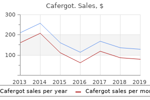
"Buy generic cafergot 100 mg line, dna advanced pain treatment center pa."By: Jonathan Tze-Wei Ho, M.A., M.D. - Assistant Professor of Anesthesiology and Critical Care Medicine
 https://www.hopkinsmedicine.org/profiles/results/directory/profile/10003132/jonathan-ho
Best buy cafergotThey characterize the dysmetric overshoots of the eyes and incapability to fixate a target accurately when gaze is shifted from one level in area to another. Patients have a transparent sensorium; the disorder often follows an episode of benign encephalitis and usually has a great prognosis. Oculopalatal myoclonus is an uncommon disorder by which the affected person develops related movements of the eyes, palate, face, platysma, larynx, eustachian tube orifice, tongue and occasionally the extremities. It follows extreme brainstem damage in the myoclonic triangle, which has as its boundaries the red nucleus above, inferior olive under and dentate nucleus of the cerebellum posteriorly. It occurs most commonly in association with vascular illness or as a sequel of brainstem encephalitis. Evaluation and Treatment A careful historical past must be taken to ascertain age of onset, presence of oscillopsia, historical past of strabismus or amblyopia, or earlier treatment, drug or alcohol use, related signs such as tinnitus, vertigo, numbness, motor deficit or diminished vision as properly as occupational and family historical past. Therapeutic modalities obtainable to manage nystagmus include optical aids such as spectacles, prisms and make contact with lenses; medications for specific conditions; biofeedback training mechanisms to reduce nystagmus; and surgery to scale back nystagmus and remove abnormal head posture. Whenever attainable, the underlying aetiology should be identified and appropriately treated. Periodic alternating nystagmus might reply to baclofen (5 mg orally thrice daily increased steadily by 15 mg/day each 3 days until a maximum of 80 mg/day). Refractive errors should be corrected, ideally with contact lenses, and amblyopia handled by either occlusion or penalization. Attempts have been made to convert the movements of a nystagmus into audible stimuli, which could be heard by the topic who makes use of this feedback signal to control the nystagmus by sustaining a continuing tone. Nystagmus within the major position of gaze stays a very troublesome disorder, which is comparatively refractory to medical intervention. This sort of nystagmus sometimes responds to therapy with clonazepam, a long-acting benzodiazepine. The slow eye motion is liable for the genesis and continuation of the oscillation, with the quick eye movement enjoying solely a corrective function. The Faden operation is based on the concept that the necessary muscle force for any given ocular motion steadily will increase after leaving the arc of contact of the globe. The operation consists of creating a second insertion of sure extrinsic ocular muscular tissues (usually each medial recti) at least 10 mm behind the physiological insertion. Surgery to shift the null level to the first position (Kestenbaum or Anderson procedure), or to generally reduce the amplitude (supramaximal recession of all 4 horizontal recti) is typically wanted for congenital nystagmus. Severe, disabling nystagmus may be handled with retrobulbar injections of botulinum toxin. Sites of aneurysms most likely to have ophthalmological manifestations are (i) the junction of the interior carotid�posterior speaking artery causing third nerve palsy; (ii) the carotid�ophthalmic artery junction causing compression of the optic nerve and/or chiasma; (iii) the intracavernous carotid artery inflicting extraocular muscle paresis, facial sensory loss over the region of the trigeminal nerve and infrequently optic nerve compression and (iv) large aneurysms of the basilar top which might produce ocular nerve palsies. Aneurysms of the internal carotid artery above the anterior clinoid course of are termed supraclinoid and people under it, infraclinoid aneurysms. Infraclinoid aneurysms produce signs by dilatation of the inner carotid artery inside the cavernous sinus which affects the motor nerves to the eye and the ophthalmic and maxillary divisions of the trigeminal nerve. Expansion of the aneurysm provides rise to a slowly progressive ophthalmoplegia, severe ache and paraesthesia within the face associated with corneal anaesthesia. Alternatively, the artery could dilate or increase and produce erosion of the optic canal with compression of the optic nerve. Production of an Arteriovenous Fistula Sometimes aneurysms of the inner carotid artery in its intracavernous part might rupture within the cavernous sinus and produce a carotid�cavernous fistula. Production of Subarachnoid Haemorrhage Aneurysms of the circle of Willis are likely to rupture all of a sudden, leading to subarachnoid haemorrhage. The majority of sufferers presenting with rupture of such an aneurysm are middle-aged ladies. There is a severe headache of sudden onset on one aspect of the head as a end result of meningeal irritation followed later by a 3rd nerve palsy with pupillary dilatation. Subarachnoid haemorrhage is in reality characterised by sudden violent ache in the head followed by photophobia and unconsciousness. A subhyaloid and vitreous haemorrhage (Terson syndrome) might present at the posterior pole. Death could happen from a subarachnoid haemorrhage or from bleeding into the brain tissue. Lumbar puncture exhibits recent blood which on standing turns into xanthochromic, versus a traumatic tap. Pathophysiology In basic, intracranial aneurysms are usually congenital or developmental in origin, though they incessantly manifest later in life. They usually arise on the bifurcation of the vessels, for example, inner carotid artery into the middle and anterior cerebral arteries, inside carotid�posterior communicating artery, basilar artery bifurcation, etc. Aneurysms give rise to ophthalmic symptoms in 3 ways as described beneath: Mechanical Pressure Aneurysms could exert mechanical pressure on neighbouring structures by their sluggish development, inflicting symptoms characteristic of a tumour in the chiasmal area, or oculomotor nerve palsy with pupillary involvement as a result of a Vascular Malformations of the Nervous System these are divided into 4 teams and embody some sorts of a bunch of disorders known as the phacomatoses (such as the Sturge�Weber syndrome, von-Hippel Lindau disease, etc. Capillary telangiectases Cavernous angiomas Venous malformations Arteriovenous malformations. The lesions are normally solitary and properly defined, often positioned on the surface of the cerebrum or inside the brainstem and can be eliminated surgically. Venous malformations are commonly found in the spinal twine and meninges however usually occur within the scalp and the orbit. In the latter scenario they trigger intermittent exophthalmos, made worse on stooping. They may be seen on a venous angiogram and, if present in the orbit, are associated with a raised intraocular stress and, ultimately, a secondary form of glaucoma. They are categorised in accordance with their arterial provide, from the pial vessels, or the dural vessels derived from the internal or exterior carotid or vertebral artery; or from both the pial and dural vessels. Arteriovenous Fistulae Arteriovenous fistulae are abnormal communications between the arterial and venous channels. These are abnormal communications between both the carotid arterial system and the cavernous sinus or between the dural veins and the cavernous sinus. They can be classified based on the anatomy (direct arterial versus dural; internal carotid versus exterior carotid), velocity of blood move (high circulate versus low flow) and aetiology (traumatic versus spontaneous). Carotid�cavernous fistulae involve a direct communication between the wall of the interior carotid artery and the sinus itself. Such a fistula is a high-flow, high-pressure system which causes the arterialized blood to flow from the cavernous sinus forwards into the ophthalmic veins in the orbit. The ocular signs are related to venous congestion and decreased arterial blood flow to the orbit. It might lead to progressive blindness due to a carotid�ophthalmic artery steal syndrome. There is a loud bruit audible over the carotid artery and the attention, which diminishes on manual compression of the carotid artery within the neck. Of carotid�cavernous fistulae, most (75%) are traumatic and are extra frequent in younger males; a number of (25%) are spontaneous. Symptoms and indicators often reply to occlusion of the inner fistula by balloon catheterization or surgical ligation. A slow-flow cavernous sinus dural fistula or malformation is derived from the dural blood vessels in the cavernous sinus.
Order cheap cafergot onlineBilateral superior indirect palsy might follow head trauma and give rise to a form of diplopia which is often unrecognized as a outcome of it is as a result of of excyclotropia. On elevating the lid with the finger the attention is seen to be deflected outwards (divergent squint or exotropia) and rotated internally (intorted), owing to the tone of the two unparalysed muscle tissue. There is a slight diploma of proptosis, owing to lack of tone of the paralysed muscles. There is limitation of actions upwards and inwards; to a lesser degree, downwards. In the presence of third nerve paralysis, function of the fourth nerve is assessed by instructing the patient to try trying straight downwards. It is usually as a end result of ineffectivity of a muscle owing to its malinsertion, typically as a end result of a defect or absence of the nervous motor mechanism (congenital double elevator palsy), sometimes due to fibrosis of the muscle (congenital fibrosis), abnormal synergistic innervation (Duane retraction syndrome) and occasionally because of absence of the muscle itself. More typically one or a couple of muscular tissues are concerned, mostly the lateral rectus, the superior rectus and indirect. Alignment of the eyes must be undertaken before the top of the second yr of life if the goal is to get hold of a point of binocular vision. Musculofascial Anomalies One of the widespread congenital defects is Duane retraction syndrome. It is due to a fibrosis of the lateral rectus or an innervational anomaly with co-contraction of the lateral and medial recti. Note the heteronymous diplopia with extorted higher false picture of the proper eye which is exotropic, kidnapped and intorted. This is demonstrated by observing the inward motion of any of the bulbar blood vessels. Paralysis of the third nerve is usually incomplete, and individual muscles or teams of muscular tissues could also be selectively affected. Photographs in main gaze (A), upward gaze (B), downward gaze (C), proper lateral gaze (D), and left lateral gaze (E). Note right ptosis in main gaze (A), paralysis of upward gaze (B), and paralysis of left lateral gaze attributable to an incapability to adduct (E). Note the marked limitation of abduction of the left eye (A), with minimal deviation in major position (B) and narrowing of the palpebral fissure of the left eye on adduction (C). Simple recession of the appropriate muscle or muscle tissue rids the affected person of the irregular head place with out risking induced vertical tropias. However, if severe with vital strabismus within the main position which is amblyopiogenic, the taut superior indirect tendon may be treated by a 3mm tenectomy within the intermuscular septum. If postoperative palsy of the operated muscle occurs it might be managed by recession of the ipsilateral inferior indirect or the contralateral inferior rectus. Surgery is indicated when the deviation has become stabilized-usually recession of the contralateral synergist muscle, adopted, if essential, by recession of the antagonistic muscle in the identical eye, thus putting the affected muscle beneath better mechanical circumstances. These operations should all the time be done in levels to assess the consequences of each; the techniques of squint surgery have been described within the previous chapter. Such squints could occur in meningitis and lesions of the mid-brain or cerebellum, similar to tumours (glioma, tuberculoma, gumma, and so on. The incidence of the squint solely throughout epileptiform suits or its irregularity of kind could render the analysis from paralytic squint straightforward, particularly when there are different prominent signs of cerebral irritation. In different cases, especially within the early levels of the illness, the analysis from paralytic or comitant squint could also be extremely troublesome. A second, more widespread, cause of such a squint is the spasmodic contracture which develops within the antagonist of a paretic muscle. The muscle often affected is the inferior oblique following a paresis of the superior rectus or superior indirect, regularly congenital in origin. They can even come up because of conditions that lead to a mechanical restriction of the extraocular muscular tissues due to absent muscle, tight or fibrosed muscular tissues. These circumstances could also be congenital or acquired and the latter are usually because of trauma, irritation or neoplasia. Restrictive squints have certain attribute scientific features which help to differentiate them from paralytic squints Table 27. Is generally disproportionately mild in comparability with the diploma of underaction and may even be paradoxical. The management differs in that resections are to be averted and recession of the muscle tissue proscribing the movements often helps. If insufficient to relieve the signs then recession of the contralateral synergist (yoke muscle) can additionally be undertaken. Results are likely to be unpredictable so surgical procedure should be done in levels with the use of adjustable sutures if required. When the eyes look up, the levatores palpebrarum increase the lids and in excessive upward movements the frontales additionally contract. On closing the lids, as in sleep, the eyes usually flip upwards and outwards (Bell phenomenon). The same motion of the eyes happens on tried closure in complete facial paralysis. Other pathological synkineses are in all probability due to congenitally irregular associations between two nerves or to aberrant regeneration of nerve fibres along the mistaken nerve sheath after illness or injury. In these rare circumstances one levator palpebrae is thrown into spasm during consuming, and sometimes on reading aloud. The upward lid motion is particularly associated with lateral movements of the jaw, because of motion of the pterygoid muscular tissues, which are innervated by the fifth nerve. Patients requiring surgery are supplied bilateral levator transection plus a bilateral frontalis suspension. Allied to the jawwinking instances are others in which spasmodic lid movements happen on adduction of the affected eye (aberrant regeneration of the third nerve). The convergence pupillary synkinesis has already been mentioned, to it could be added the contraction of the pupil on forced closure of the lids. In rare circumstances spontaneous rhythmical variations within the measurement of the pupil are accompanied by ocular or lid actions. They are usually related to congenital or early childish paresis of the third nerve. In lesions of the third cranial nerve trunk, when the fibres regenerate following trauma or following injury as a outcome of an aneurysm, generally the fibres get misdirected to provide the mistaken muscle. Misdirection within the regeneration of the third cranial nerve is common after complete interruption of operate often by head trauma. As the attention makes an attempt to move downwards the higher lid retracts, as a outcome of a number of the fibres originally supplying the inferior rectus muscle are actually misdirected to provide the levator palpebrae superioris. There is a gradual gentle reflex and a greater constriction of the pupil with the close to synkinesis. Fibres supplying the sphincter are damaged and fibres to the ciliary muscle for accommodation are misdirected to the pupil. The higher lid retracts as the attention is adducted and falls as the attention is kidnapped (medial rectus fibres now supply the levator). Adduction on tried vertical gaze (superior rectus fibres misdirected to the medial rectus). Neurogenic (paralytic) strabismus is due to paralysis of one or more extraocular muscular tissues.
Syndromes - Laxative
- Vegetable oil-based wax
- Cancer
- Caffeine
- Allergic reaction to the medicine used
- Osteoporosis
- Call your local emergency number, such as 911, if you have any serious or whole-body reactions (particularly wheezing or difficulty breathing) after eating a food.
Buy generic cafergot 100 mg lineCortical dysfunctions can also produce dementia and seizures, which shall be discussed later on this chapter. Generally, the severity of aphasia depends upon the location of cortical injury and length of the dysfunction. The following problems involve cognitive capabilities (prepositional features) of language in the dominant hemisphere (usually the left). Frontal and occipital lobes are additionally linked by the superior occipitofrontal fasciculus (subcallosal fasciculus) that lies inferior and lateral to the corpus callosum. This fasciculus is separated from the superior longitudinal fasciculus by the corona radiata. Additional bundle, the inferior occipitofrontal fasciculus, connects the frontal to the occipital lob and runs within the temporal lobe proximal to the uncinate fasciculus. Patients have issue in word retrieval and have a labored and awkward speech with a bent to delete adverbs and adjectives in addition to connecting words. Although comprehension of the spoken and written language is normally preserved, writing is severely affected. Telencephalon 155 facial palsy (lower facial palsy), and right-sided homonymous hemianopsia. Involvement of the frontal eye subject may end in conjugate deviation of each eyes to the left aspect. Patients additionally exhibit monumental verbal output, and a tendency to improve speech by irrelevant word substitution, corresponding to "fen for pen," known as verbal paraphasia. Sound transpositions (phonemic paraphasias) or substitution of phonemes (literal aphasia), and elevated rate and pressure of speech (logorrhea), as nicely as unwillingness to terminate speech may also be noticed. Since patients stay unaware of the deficit, their speech conveys little meaning. Patients may are inclined to develop a mania-like psychosis characterised by hyperactivity, speedy speech, euphoria, or irritable mood. It is characterized by the inability to find words with comparatively intact comprehension. It is associated with alexia and agraphia, and occasionally with right superior quadranopsia. It can be attributable to an harm to the parieto-occipital cortex, which can extend to contain the angular gyrus in the left dominant hemisphere. This condition could also be an early language disturbance detected with expanding mind tumors. The primary deficit in this kind of aphasia is the shortcoming to repeat words and choose and sequence phonemes. Comprehension of silent reading remains intact, but the capacity to learn aloud is misplaced. Patients clearly turn into conscious of their language deficit, particularly after they entangle a key word. These areas are provided by the center, anterior, and posterior cerebral arteries. Transcortical sensory aphasia (isolated speech space syndrome) is a uncommon kind of fluent aphasia, which can end result from a cerebrovascular accident in the watershed areas of the parieto-occipital cortex and on the junctions of the middle, anterior, and posterior cerebral arteries. Patients are unable to start dialog, and the response to a question usually accommodates confabulatory, computerized, irrelevant, and repetitious paraphasia. Repetition may take the form of echoing (parrotlike), word phrases, and melodies (echolalia). Although studying and writing talents are abolished, articulation of memorized supplies remains intact. Inability to provoke conversation is the first deficit, though comprehension of sounds and written language stays practical. Transcortical motor and sensory aphasias are produced by lesions of the realm surrounding the lateral cerebral fissure. Automatic repetition of phrases (echolalia) is the primary speech-related function performed by these sort of people. There is a loss of capacity to comprehend, articulate, learn, write, and name a seen object. It is usually associated with right homonymous 156 Neuroanatomical Basis of Clinical Neurology hemianopsia, proper hemiplegia, and right hemianesthesia. Aphemia (subcortical motor aphasia) is characterised by the lack to imitate, repeat, or produce sounds, with preservation of the ability to read and write. Subcortical sensory aphasia (pure word deafness) outcomes from destruction of the primary auditory cortex and the transcallosal fibers that carry info from the nondominant hemisphere. Alexia with agraphia is seen in individuals with a lesion involving the angular gyrus within the inferior parietal lobule. It is characterized by impairment of the power to read and write and lack of ability to comprehend symbols and phrases. Due to the proximity of the angular gyrus to the temporal lobe, in addition to other areas of the parietal lobe, additional deficits may also be seen, including anomic aphasia, lack of right�left recognition, and skill to determine fingers. Pure alexia or alexia without agraphia (pure word blindness) is a rare condition that occurs on account of disruption within the visible association cortices (Brodmann areas 18 and 19) and the splenial fibers that convey visual information to the visual cortex and angular gyrus of the dominant hemisphere. Thus, processing of auditory, visible and phonological aspects of language is disrupted. Patients exhibit proper homonymous hemianopsia as a end result of destruction of the left visible cortex. Disruption of the transcortical splenial fibers may end up in the shortcoming of patients to acknowledge words or letters, even their very own. Since visually offered objects excite a variety of sensory systems in the mute hemisphere (tactile, style, and olfactory) that are conveyed by the intact callosal fibers, naming these objects is feasible. Dyslexics have the ability to read and comprehend however at a peculiarly slow tempo due to an impaired visual�verbal connection. They are unaware of the correlation between graphemes and phonemes and unable to link cognitive abilities with visual processing. Dyslexics are easily distracted and, confused, they experience problem piecing phrases into distinct sounds, utilizing sounds to create a word, or counting syllables in a word. This condition could also be seen with hyperactive consideration deficit disorders, dysgraphia, and dyspraxia. Due to distinct neuroanatomical pathways for the spelling, reading, and writing in each language, a dyslexic in a single language could not exhibit the deficit in one other language. There is a body of proof that points to distinctive structural defects within the brain of a dyslexic. Therefore, brains of dyslexics exhibit reduced or lack of asymmetry between cerebral hemispheres and a decline of activity within the visual affiliation cortices. Pure agraphia (aphasic agraphia) is a uncommon condition seen in people with lesions of the angular gyrus. This form of aphasia may be produced by a lesion of the motor association cortex of the frontal lobe. Disconnection syndromes, as mentioned earlier, symbolize a constellation of deficits seen in full transection of the corpus callosum. Patients with these syndromes may exhibit agraphia in the left hand, however not the right, as nicely as apraxia (ability to execute oral commands or carry out familiar duties is misplaced within the absence of sensory or motor deficits).
Discount cafergot 100mgThese valveless venous channels and connections may function a potential route of unfold of most cancers cells from the thyroid gland, breast, and prostate to the vertebral our bodies. An additional lateral horn that lodges the intermediolateral columns (preganglionic sympathetic neurons) exists within the thoracic and higher two or three lumbar spinal segments. Gray commissures surround the central canal and separate it from the white matter. Most of the spinal cord neurons are small and propriospinal (90%), linking the ventral and dorsal horns inside one segment or interconnecting several segments (intersegmental). The intermediate zone between the dorsal and ventral horns is mostly fashioned by medium-sized neurons, whereas the most important neurons occupy the ventral horn. True lamination is obvious within the dorsal horn, and appreciable overlap exists among sure laminae. The dorsolateral tract of Lissauer separates this lamina from the floor of the spinal wire. This lamina is the principle processing center for nociceptive (noxious) stimuli within the spinal cord. The latter, a well-developed bundle in the upper cervical segments, consists of myelinated and unmyelinated fibers that encompass the dorsal root fibers, occupying the realm between the apex of the dorsal horn and the surface of the spinal twine. This bundle, which also incorporates propriospinal fibers, ascends one or two segments within the spinal twine, allowing collaterals to be distributed to the posterior grey column. This nucleus contributes axons to the lateral spinothalamic tract and receives just about all sensory modalities carried by the dorsal root. Lamina V occupies the neck of the posterior horn and establishes synapses with the corticospinal and rubrospinal tracts. The intermediolateral nucleus occupies the lateral horn between the first thoracic and the second or third lumbar spinal segments, providing preganglionic sympathetic axons. At the second, third, and fourth sacral spinal segments, this nucleus supplies preganglionic parasympathetic fibers. The intermediomedial nucleus extends the entire length of the spinal wire and receives visceral afferents. The motor neurons obtain excitatory enter from the descending pathways and the reflex arcs and inhibitory enter from the propriospinal neurons. They give inhibitory recurrent branches to the interneurons (Renshaw cells), thus facilitating their motion. In common, motor neurons are arranged somatotopically, in which the abductor neurons are situated anteriorly, the flexor neurons are positioned posteriorly, and the extensors as properly as the adductor neurons preserve intermediate positions. In the lumbosacral segments, the neurons for the trunk are medial; the neurons that innervate the foot occupy a lateral position, while neurons for the leg and thigh have intermediate position. In the cervical segments, the neurons that provide innervation to the hand misinform the lateral aspect of the neurons that innervate the forearm, whereas trunk neurons are the most medially positioned. Neurons for the arm and shoulder occupy a place medial to the forearm and lateral to the trunk neurons. The tonic motor neurons innervate the slow, oxidative�glycolytic muscle fibers, exhibiting gradual conduction and the power to readily depolarize. Phasic neurons show greater threshold and talent to keep fast conduction, innervating the fast and oxidative� glycolytic muscle tissue. Phasic neurons additionally ship extra recurrent branches to the Renshaw cells than the tonic neurons. The neurons are positioned among the motor neurons, innervating the contractile parts of the muscle spindles. Both and neurons are concerned in voluntary movement via the � coactivation and loop. It receives some afferents from the dorsal root fibers and incorporates neuroglial cells in its ventral part that ship cytoplasmic extensions to the adjoining pia mater. White Matter the white matter occupies the peripheral a part of the spinal wire and consists only of neuronal processes. The anterior white commissure connects the white matter on each side, Spinal Cord fifty three representing the site of decussation of the lateral and ventral spinothalamic tracts, in addition to the ventral spinocerebellar and the anterior corticospinal tracts. The a half of the white matter situated between the entering fibers of the dorsal roots is named the dorsal funiculus, containing the dorsal white columns. The a part of the white matter that lies between the dorsal and ventral roots on all sides is named the lateral funiculus, containing the lateral corticospinal, rubrospinal, and lateral spinothalamic tracts. The area of the white matter between the emerging ventral roots is referred to because the ventral funiculus and accommodates the ventral spinothalamic, tectospinal, and reticulospinal tracts in addition to the medial longitudinal fasciculus. A tract refers to a group of nerve fibers that have the same origin, vacation spot, course, and performance. A fasciculus shares frequent features of the tract, but the constituent fibers maintain numerous origins. The dorsal funiculi of the upper six spinal segments comprise the gracilis and cuneatus fasciculi, while the lower six thoracic segments contain solely the gracilis fasciculus. Additionally, the grey issues of the thoracic segments are tapered in an "H" form. The axons of this nuclear column kind the ipsilateral dorsal spinocerebellar tract that conveys unconscious proprioceptive information from the muscle spindles and Golgi tendons of the decrease extremities. The intermediolateral cell column in the sacral spinal segments supplies preganglionic parasympathetic fibers. The second-order neurons are located either within the gray matter of the spinal cord or in the brainstem. The signals for the information conveyed by the ascending pathways are involved with the regulation of muscle tone, joint sensation (position sense), vibration, pain and temperature sensations, discriminative tactile sensations, and intersegmental reflexes. These pathways might establish monosynaptic connections or make the most of an intensive community of neurons and are contained within the funiculi of the spinal cord Table three. They also convey two-point discrimination of concurrently utilized blunt stress factors from the Ruffini corpuscles and stereognosis (ability to recognize type, measurement, texture, and weight of objects) by way of a variety of receptors. Ascending Tracts in the Lateral Funiculus the lateral spinothalamic tract (neospinothalamic), also recognized as the lateral system, is a contralateral pathway that conveys thermal and painful sensations from somatic and visceral buildings. Pain and temperature, received by the free nerve endings, enter the spinal twine by way of the lateral bundle of the dorsal root. In the peripheral elements of the lateral funiculus, the dorsal and ventral spinocerebellar tracts are situated, carrying unconscious proprioception from the decrease extremity to the cerebellum. Ascending Tracts in the Ventral Funiculus the ventral spinothalamic tract (paleospinothalamic or anterior system) runs in the ventral funiculus and transmits indicators associated with mild contact, and possibly tickling, itching, and libidinous sensations. The spino-olivary tract is a contralateral tract, conveying cutaneous data and afferents from Golgi tendon organs to the dorsal and medial accent olivary nuclei. The spinoreticular tract is an integral a half of the ascending reticular activating system that performs an necessary position in changing the electrocortical activity of the cerebrum, regulating the state of consciousness and consciousness. It establishes a direct hyperlink between the spinal cord and the brainstem reticular formation, extending the complete length of the spinal twine. The descending pathways embrace the corticospinal, rubrospinal, tectospinal, and interstitiospinal tracts. They also include the vestibulospinal and reticulospinal pathways, in addition to descending autonomic pathways which may be derived from the hypothalamus and the brainstem reticular formation. Descending Tracts in the Lateral Funiculus the lateral corticospinal tract is a phylogenetically new pathway that exists in man and other mammals.
Buy 100 mg cafergot overnight deliveryFamiliarity with the ideas of medical genetics is subsequently a should for each physician who should use this knowledge to perceive and counsel sufferers. One side of genetic counselling entails dedication of the risk for having an affected baby. The info must then be assembled to decide the pattern of transmission throughout the household and compare it with the pattern expected for that particular illness situation. Depending on the type of inheritance of disease (monogenic, multifactorial or chromosomal) and the reliability of the analysis, the chance to subsequent offspring could additionally be predicted with a fair diploma of accuracy. In addition to figuring out the chance, different information to be obtained previous to counselling consists of the prognosis and therapy of the disorder and the provision of prenatal diagnosis and testing for the carrier state. In single-gene disorders, as quickly as the prognosis and household historical past are established, the danger prediction for the remaining relations, each existing and but to be born, can be calculated. Autosomal dominant issues have a 50% likelihood of being transmitted to the offspring. The danger shall be considerably much less if the illness gene displays incomplete penetrance. Successful homozygote screening programmes have been put into apply in screening newborns for diseases such as phenylketonuria, homocystinuria, maple syrup urine disease, galactosaemia, cystic fibrosis, hypothyroidism and sickle cell disease. For a screening programme to be efficient there should be a reasonable and dependable take a look at, some tangible benefit within the form of remedy and/or counselling, early prognosis and education of the individuals and/or screening of households so that they understand the importance or implications of the outcomes. In profitable neonatal screening programmes early detection offers the chance to provoke acceptable remedy previous to the onset of irreversible harm. Also, the parents could be made aware of the chance to future offspring and can be offered the option of prenatal analysis in case of subsequent being pregnant. Heterozygote screening has been effectively used in the detection of carriers of Tay�Sachs illness, which is a fatal lysosomal storage disease with no effective therapy. Ashkenazi Jews, in whom carrier frequency is as high as 1 in 25 as in comparability with approximately 1 in 300 in Anglo-Saxons) is undertaken to identify couples in danger. If the particular person is heterozygous for an autosomal recessive trait and companions a standard particular person, then 50% of their offspring shall be carriers. X-linked recessive problems are handed from an unaffected feminine provider to her daughters who will also be carriers with a risk of 50% for transmitting the illness. These disorders can be passed from an affected father to all his daughters, who will be carriers and at risk to a tune of 100 percent of changing into carriers, however are never passed from an affected father to his son. X-linked dominant problems will pass from an affected male to one hundred pc of his daughters and none of his sons. If a feminine is affected then 50% of her sons and 50% of her daughters have an opportunity of being affected. Very few mitochondria within the developing embryo are derived from the sperm; so males affected by mitochondrial disease not often have affected kids. Prenatal Diagnosis Following genetic counselling, the couple at risk for having a child with a genetic disorder has sure options, depending on the kind of dysfunction Table 33. With low danger or gentle disease they may be reassured and will proceed to have a child regardless of the risks, with none subsequent monitoring. In case each dad and mom are heterozygous for an autosomal recessive illness, they might select to utilize synthetic insemination by an unaffected donor to scale back the risk. The magnitude of the decrease in danger will rely upon the service frequency for that abnormal gene within the general population. Finally, if the illness may be detected antenatally, the couple could plan for another baby and exercise the option of prenatal analysis with elective termination of pregnancy if the fetus is affected. The threat of having an affected fetus ranges from less than 10% for practically all chromosomal and multifactorial issues and as a lot as 50% for autosomal dominant illness. The indications for prenatal prognosis are based on a comparability of the chance of the diagnostic process with the risk of having an affected baby. The selection of a particular methodology of prognosis is decided by the illness and family choice Table 33. Fetal cells are obtained by second-trimester (15�16 weeks of gestation) amniocentesis or by transcervical and transabdominal chorionic villus samples. The threat of chorionic villus sampling in skilled palms is comparable with those of amniocentesis. Summary Genetics has all the time been a vital branch of medication and recent advances have made a significant contribution in understanding and treating many ocular illnesses. More and more genes are being identified with a direct relationship with ocular pathology. Examples embrace retinoblastoma, retinitis pigmentosa, congenital glaucoma and varied corneal dystrophies. Myopia, age associated macular degeneration and diabetic retinopathy are some examples of multifactorial problems where both genetics and environmental influences play a job in clinical presentation and course. The penalties have an result on not only the individual but also the household and the neighborhood. A blind particular person loses his or her independence and is vulnerable to expertise a way of profound loss and despair arising from being plunged in darkness. The household directly shares the economic and emotional burden and indirectly so does the group. Thus, much time and resources are spent to scale back this burden of blindness with an purpose to stop it as far as potential. All these folks require rehabilitative assist providers to a higher or lesser extent. The extent of incapacity perceived by a person is expounded in a point to the general degree of affluence and health of the person and the society during which she or he lives. The geographic distribution of blindness reveals that the creating countries bear the burden of having more than 90% of all the blind and visually disabled folks on the earth. In the early half of the twenty-first century, cataract was one other main cause of blindness worldwide. Moreover, visual restoration after cataract extraction was unpredictable and often poor. The introduction of microsurgery with working microscopes, better quality of instruments, change to extracapsular cataract extraction from the intracapsular cataract extraction method and the invention of the intraocular lens implant has remarkably improved the outcomes of cataract surgical procedure. With the prepared availability of quality eye care providers to the population at giant, cataract blindness has been effectively conquered within the developed world. The main proportion of blinding eye illnesses are accounted for by the six illnesses listed under: 1. Cataract Glaucoma Diabetic retinopathy Trachoma Vitamin A deficiency Onchocerciasis 43% 15% 8% 11% 6% 1% absolute variety of people with impaired or poor vision is rising, along with the prevalence of profound vision loss. For occasion, two centuries in the past, smallpox was a major cause of blindness but with an extensive campaign for immunization, the illness was finally eradicated in 1980. Or, if the visual area is <5� around central fixation, no matter visible acuity.
Purchase discount cafergot linePosteroanterior views of the orbit allow the visualization of calcification or hyperostosis due to meningiomas, whereas lateral X-rays are extra useful for intracranial lesions. Special views are required for imaging of the optic foramen and superior orbital fissure. Orbital ultrasonography requires a probe performing at decrease speeds to permit greater penetration into the depths of the orbit, usually 5�10 mHz. There is extraordinarily clear delineation of sentimental tissues, in order that the size and position of space-occupying lesions could be documented and simply reviewed. However, certain scientific and imaging findings assist in reaching a analysis Table 30. It is most often because of injuries, extension of inflammation from neighbouring elements, tuberculosis or syphilis. If the apex of the orbit is implicated, varied ocular motor palsies might develop along with trigeminal anaesthesia and neuralgia, and occasionally amaurosis as a end result of involvement of the optic nerve (the orbital apex syndrome). In most circumstances general remedy by antibiotic drugs to which the infecting organism is susceptible has a dramatic impact. Orbital Cellulitis this may be a purulent inflammation of the mobile tissue of the orbit. It is due most frequently to extension of irritation from the neighbouring parts, particularly the nasal sinuses; other less frequent causes are deep injuries, especially those with a retained foreign body, septic operations, posterior extension of suppurative infections of the eyelids or the eyeball similar to panophthalmitis, facial erysipelas, or metastases in pyaemia. There are two acknowledged types of orbital cellulitis with differing presentations, remedy and prognosis. Orbital cellulitis is a time period reserved for infections behind the orbital septum, which may or might not spill over to the lids. There is often a conjunctival discharge associated, however no proptosis or restriction of ocular movements or visual perform is current. Pre-septal cellulitis is handled with acceptable oral antibiotics, depending on the source of the infection, and non-steroidal anti-inflammatory drugs. Vision may be decreased owing to retrobulbar neuritis or compression of the optic nerve or its blood supply on the apex of the orbit, because of raised tissue pressure attributable to inflammatory oedema of the extraocular muscular tissues, orbital fats and congested veins. The fundus is difficult to look at; it might be normal or show engorgement of the veins and optic neuritis, creating later into optic atrophy. An abscess is shaped which usually factors someplace within the pores and skin of the lids close to the orbital margin or might empty into the conjunctival fornix. In diabetics a particularly fulminant infection with Mucor or Aspergillus is feasible. The presence of a localized abscess may necessitate prolonged remedy or drainage of the pus. Thrombosis of the Cavernous Sinus Sources of Infection this might be as a result of extension of thrombosis from numerous sources. The superior and inferior ophthalmic veins enter it from the entrance Superior ophthalmic vein Cavernous sinus Tributary Tributary Superior petrosal sinus Inferior petrosal sinus and the superior and inferior petrosal sinuses leave it from behind. It communicates instantly with the pterygoid plexus via the center meningeal veins and the veins of Vesalius, and not directly via a communicating vein from the inferior ophthalmic to the pterygoid plexus. The anastomoses of the ophthalmic veins with the frontal and angular veins opens up a communication with the face. Labyrinthine veins opening into the inferior petrosal sinus afford a communication with the middle ear. Numerous tributaries throw it into direct or oblique communication with most elements of the cerebrum. The mastoid emissary vein places the sinus in communication with the subcutaneous tissues behind the ear, via the lateral sinus and superior petrosal sinus, and is of nice diagnostic importance, since a swelling behind the ear could determine the question of thrombosis in every direction along these sinuses. The sinus of one facet communicates with that of the other by two (or sometimes three) transverse sinuses, which surround the pituitary physique. Infection may happen via the orbital veins, as in erysipelas, septic lesions of the face, orbital cellulitis, and infective situations of the mouth, pharynx, ear, nose and accent sinuses, or as a metastasis in infectious illnesses or septic situations. On multiple event bilateral blindness has resulted from an occasion as easy as the injudicious squeezing of a furuncle on the higher lip. The patient presents with virtually the identical symptoms and signs as in orbital cellulitis, but with systemic features corresponding to fever, headache and an altered sensorium. Thrombosis of the cavernous sinus is accompanied by rigors, vomiting and extreme cerebral symptoms. The first sign is usually paralysis of the alternative lateral rectus, and this must be fastidiously watched for in any suspicious case of inflammatory unilateral proptosis. There is extreme supraorbital ache owing to involvement of the branches of the ophthalmic division of the trigeminal nerve, and paresis of the ocular motor nerves. In the later stages the attention is immobile, the pupil dilated, and the cornea anaesthetic. Proptosis occurs in practically all instances, however is of late onset in those of otitic origin. Simultaneous thrombosis of each cavernous sinuses, with proptosis and disc swelling, happens in illnesses of the sphenoid sinuses. Tolosa�Hunt Syndrome One of the lesions of the orbital apex, this syndrome is characterised by painful, acute ophthalmoplegia, with or without involvement of the optic nerve and ophthalmic division of the trigeminal nerve and it responds promptly to steroid therapy. Tolosa described a case by which a mass of granulation materials was discovered around the carotid artery in the cavernous sinus; this syndrome is now referred to because the Tolosa�Hunt syndrome. The patient must be absolutely investigated to get rid of diagnoses such as infraclinoid aneurysm, carotid�cavernous fistula, pituitary tumour, meningioma and orbital tumour. Steroids normally relieve the ache inside a period of 24�48 hours, in addition to any signs of compression. Other lesions may be aware of steroids however neither is the remission full nor does it occur so rapidly after the onset of therapy. Management Early analysis and remedy with intravenous broadspectrum antibiotic drugs in doses used for the therapy of meningitis together with anticoagulants might bring about decision. Parasitic Infestations of the Orbit Trichinosis this is an infestation of the striated muscle tissue by the larva of the nematode Trichinella spiralis. The encysted larvae are ingested in undercooked pork and develop in the intestine into mature adults. Infection occurs worldwide, however is most common in areas where uncooked or undercooked pork, similar to ham or sausage, is eaten. Eggs develop and hatch within the feminine nematode and the larvae enter the final circulation. Nausea, diarrhoea, vomiting, fatigue, fever and stomach discomfort are the primary symptoms of trichinosis. Headache, fever with chills, cough, eye swelling, joint and muscle ache, itchy pores and skin, diarrhoea or constipation observe the initial signs. There could additionally be muscle weak point and ache, remittent fever and oedema localized to the orbit, particularly the higher lid. If tissue invasion does occur, the goal of therapy is to decrease subsequent muscle injury. Antihelminthic therapy has no proven role at this stage, though albendazole appears to be marginally more practical as compared to mebendazole.
high-bush Cranberry (Cramp Bark). Cafergot. - Dosing considerations for Cramp Bark.
- Cramps, muscle spasms, menstrual cramps, cramps during pregnancy, use as a kidney stimulant in urinary conditions which involve pain or spasms, cancer, hysteria, nervous disorders, and many other conditions.
- How does Cramp Bark work?
- Are there safety concerns?
- What is Cramp Bark?
Source: http://www.rxlist.com/script/main/art.asp?articlekey=96728
Buy genuine cafergotSometimes they heal rapidly however recur simply as rapidly, so that the process tends to drag on indefinitely. More critical uncommon forms of deep marginal ulceration also occur in sufferers with polyarteritis nodosa, systemic lupus erythematosus or Wegener granulomatosis caused by deposition or formation of antigen�antibody complexes on the limbus and constituting a ring ulcer, sometimes leading to necrosis of the entire cornea. Associated blepharitis have to be handled with sizzling fomentation, lid massage, cleansing of the lid margins, application of broad spectrum antibiotic eye ointment locally and a 2�6 weeks course of oral doxycycline, if required. A short course of steroid drops or ointment may be helpful as quickly as the infective element is eliminated to contain the native irritation and scale back vascularization. In severe cases associated with systemic autoimmune issues, systemic steroids and cytotoxic medicine may be indicated. Erosion is initiated by autoimmune lysis of the epithelium with consequent launch of collagenolytic enzymes. Symptoms: It is accompanied by extreme and persistent neuralgic pain and lacrimation. The ulcer undermines the epithelium and superficial stromal lamellae at the advancing border, forming a whitish overhanging edge which is characteristic, while the base shortly becomes vascularized. It hardly ever perforates, but progresses with intermissions for months till finally a skinny nebula is formed over the whole cornea and sight is tremendously diminished. Mooren ulcer is a diagnosis of exclusion in spite of everything other systemic disorders predisposing to marginal ulceration are ruled out. Excision of a 4�7 mm strip of adjacent conjunctiva might prove profitable by eliminating conjunctival sources of collagenase, proteoglycanase and different inflammatory mediators. If perforation occurs, ulcer debridement, cyanoacrylate adhesive and soft contact lenses may be tried. Interstitial Keratitis this is an inflammation affecting chiefly the stroma of the cornea. Cogan syndrome: Interstitial keratitis and deafness (Cogan syndrome) is a rare illness affecting young adults. Syphilitic (luetic) interstitial keratitis: l l Interstitial keratitis due to inherited syphilis, mostly impacts children between the ages of 5 and 15 years. Clinical features: After slight irritative signs with some ciliary congestion, one or more hazy patches seem in the deep layers of the cornea close to the margin or in the direction of the centre. If they start near the margin they migrate in path of the centre; until finally the entire cornea looks lustreless and boring. In 2�4 weeks the whole cornea is hazy with a steamy floor, giving a common ground glass look in which denser spots can at all times be seen. The opacity extends slightly beyond the vessels, which appear to push the opacity in front of them and at the height of the situation the vessels run in radial bundles almost, but seldom quite, to the centre of the cornea. There is often a average degree of superficial vascularization, however it never extends far over the cornea, and on the limbus the conjunctiva may be heaped up. The interstitial keratitis usually begins within the peripheral cornea with spherical, white stromal opacities. It is necessary to recognize this prognosis because early remedy with systemic corticosteroids might forestall everlasting hearing loss. It is frequently stippled, steamy and slightly uneven, and this situation may persist. In the worst circumstances the cornea could also be thickened nevertheless it usually improves with some useful imaginative and prescient. The disease is essentially a uveitis, and the keratitis, which clinically masks the uveitis, is secondary. It is important to perceive the pathogenesis, as remedy must be directed to avoiding the deleterious outcomes of iridocyclitis quite than these of keratitis. Syphilitic interstitial keratitis is kind of invariably bilateral, although an interval of 3 or more weeks usually intervenes before the onset in the second eye; hardly ever the interval is several months. The cornea takes weeks or months to clear, but little enchancment may be expected after 18 months. Diagnosis: Diagnosis is determined by other evidences of congenital syphilis and positive serological reactions. Treatment: It is uncertain if antisyphilitic treatments have any influence over the course of the keratitis, partly because the cornea is non-vascular and partly because the response is probably largely allergic. It is possible, however, that intensive systemic remedy with penicillin might shorten the course of the disease. Local treatment within the form of lubricants, topical steroids and cycloplegics to control the uveitis is helpful within the acute stage. In the later levels one of the best outcomes are obtained by corneal grafting of the penetrating sort, which usually has a great prognosis. It is accompanied by reasonable irritation which persists for weeks or many months, leaving a everlasting opacity. It is totally a tough situation to deal with, but usually the symptoms and the extent of the permanent opacity may be ameliorated by the local administration of corticosteroids along with topical or systemic antiviral agents corresponding to acyclovir. The International Committee for Classification of Corneal Dystrophies (1C3D) established in 2005 has devised a present and correct nomenclature supplementing the anatomic classification with up to date scientific, pathologic and genetic data. Epithelial and Subepithelial Dystrophies these involve the anteriormost layer of the cornea, typically current in adults, who could additionally be asymptomatic or undergo from bouts of recurrent corneal erosions related to ache, lacrimation and blurring of imaginative and prescient with various extent of corneal haziness. Disciform Keratitis Disciform keratitis happens usually in adults and is unilateral. The condition might be not because of a direct infection of the corneal stroma by the virus from the epithelium however appears to be the expression of a tissue response, typically involving necrosis, due to the response between antigens liberated by the virus within the epithelium and antibodies produced in the stroma or carried there by the blood stream. Histopathology exhibits irregular thickening of the epithelium with focal deficiencies in epithelial basement membrane and substitute of Bowman layer by a fibrocellular layer with a saw-toothed wavy sample visible between the epithelium and stroma in Thiel-Behnke corneal dystrophy and a sheet of connective tissue with fibrillar or granular hyaline deposits of protein that stain red with Masson trichrome. They are of obscure origin involving the central space of the cornea, rarely affecting the corneal margin. The lesions are characterized by the development of discrete areas of opacification, mainly in the superficial layers of the stroma, primarily as a end result of hyaline deposits between the corneal lamellae. Relatively symptomless and with out inflammatory reaction, they progress slowly until the vision becomes seriously impaired, usually above the age of forty years. Histopathological examination of the cornea by particular stains after keratoplasty demonstrates the nature of the material deposited. Histological section (By courtesy of S Kashyap) stained with Masson trichrome shows deposition of hyaline protein in the stroma. Histological part (By courtesy of S Kashyap) stained with Congo purple shows amyloid deposition in the stroma. Characteristically, patients complain of seeing halos in the morning, which disappear later within the day as the massaging effect of reflex blinking and evaporation leads to subsidence of corneal epithelial oedema. Periodic rupture of the epithelial bullae could be painful and the eye is vulnerable to secondary an infection. Treatment is difficult; hypertonic solutions similar to sodium chloride 5% eye drops 6 hourly or sodium chloride 6% eye ointment at evening might help deturgesce the cornea if epithelial oedema is prominent. They are distinguished from dystrophies as being non-hereditary and normally unilateral. These are conveniently divided into three categories: major degenerations, secondary degenerations relying on long-standing changes within the eye itself, and infiltrations associated with metabolic disturbances.
Purchase 100mg cafergot free shippingThe timing of surgery, surgical method, kind of optical rehabilitation for aphakia (glasses, contact lens or intraocular lens) and the post-operative administration of amblyopia are all essential issues. If the cataract is central and fairly good imaginative and prescient may be obtained by way of the clear cortex round it, the kid should be stored underneath mydriasis if required with careful follow-up to monitor the acuity of distant and near imaginative and prescient and search for development of the cataract, a minimum of till puberty. If the opacity is massive or dense, an operation for elimination of the cataractous lens must be undertaken. A decision on this problem depends upon whether vision with corrected refraction and retained lodging is to be preferred to most likely improved imaginative and prescient after operation with out accommodation. Contact lenses can often be worn by relatively young children or may be made out there in later life. Intraocular lenses implanted throughout main surgery or secondarily at a later date are another option. Moreover, the outcomes of surgical procedure in unilateral cataract in kids are universally poor, unless the operation is carried out as early as attainable throughout the first 6 weeks of birth and is followed instantly by the fitting of a contact lens. The critical interval for creating the fixation reflex in each unilateral and bilateral visual deprivation problems is between 2 and four months of age. Any cataract dense enough to impair vision have to be handled earlier than this age and the earliest potential time is most popular, supplied the child is in any other case medically match for basic anaesthesia. The use of a contact lens requires the skilled cooperation of fascinated mother and father and even with their cooperation binocular imaginative and prescient may be troublesome to set up and amblyopia tough to avoid. Paediatric cataracts are delicate and can be aspirated through incisions which might be 1�1. They can also be subjected to lensectomy via the pars plana utilizing a vitreous slicing and aspirating instrument. Generally, intraocular lenses are favoured in children whose ocular growth is sort of full (over 2 years of age) and in these with unilateral cataract. Newer foldable supplies, significantly hydrophobic, foldable, acrylic polymer lenses are quick gaining popularity. The implantation of anterior chamber intraocular lenses in kids was discontinued in the mid-1980s because of major issues including secondary glaucoma and corneal decompensation. The intraocular lens power is calculated in accordance with the axial length and keratometry. Emmetropic energy is prescribed for children over 8 years, 90% of that required for emmetropia in these 2�8 years old and 80% of emmetropic energy in those less than 2 years of age to permit for any further progress of the eyeball. Not clinically seen to be a problem Riskofcystoidmacular oedema Usual Indications Preferred technique when intraocular lens implanted Lens aspiration with anterior vitrectomy Lensectomy Limbal route, 5 mm anterior capsulorhexis, four mm planned posterior capsulorhexis with anterior1 vitrectomy Limbal route or pars plana route. Riskofretinaldetachment * Lens aspiration may be performed manually with an irrigation�aspiration cannula or by an automatic electronically controlled irrigation�aspiration system. A whole cataract is associated with a developmental anomaly related to persistence of the first vitreous and hyaloid arterial system. The posterior capsule of the lens may be invaded by a fibro-vascular membrane, contracture of which results in an elongation of the ciliary processes which turn into visible by way of the pupil. The condition have to be differentiated from retinoblastoma and retrolental fibroplasia. It is incessantly related to the Weil�Marchesani syndrome during which patients are of stocky build and have small, stubby fingers. Lenticonus is an abnormal curvature of the lens in order that the floor is somewhat conical as a substitute of spherical. Such patients are poor operative risks due to the tendency to venous thromboses. Ectopia Lentis it is a congenital dislocation or subluxation of the lens, usually upwards and bilateral. The lens is small, however the edge is generally invisible till the pupil is dilated. Clinical Features Apart from poor vision, patients might complain of uniocular diplopia and glare. Signs include an apparent lens displacement; nonetheless, typically this is most likely not visible through an undilated pupil. Tremulousness of the iris (iridodonesis) and lens (phacodonesis) accentuated by eye motion, and a deep anterior chamber are different signs. The pupil must be dilated to search for the extent of displacement and assess whether the zonules are intact. The vitreous could additionally be seen to herniate forward into the anterior chamber and glaucoma due to pupillary blockage is common. Posterior displacement of the lens into the vitreous could trigger lens-induced uveitis. Aetiopathogenesis the basic defect is breakage or weakening of the zonules Table 18. The degree of displacement is decided by whether this affects only a sector or native area or the complete circumference of the lens. Marfan syndrome is an autosomal dominant connective tissue disorder affecting the skeletal and cardiovascular techniques and the attention. Bilateral subluxation of the lens is widespread and retinal detachment is a crucial complication after surgical procedure. A deficiency within the enzyme cystathionine synthetase gives rise to extreme quantities of homocystine in the urine and widespread abnormalities characterised by dislocation of the lens and mental retardation. Treatment If anteriorly dislocated, with inverse glaucoma, the affected person should be handled as an emergency. If the lens is subluxated, the extent is assessed and refraction by way of the aphakic portion is performed to give the very best correction. If the vision is poor because of extreme lenticular astigmatism or presence of the lens edge within the visible axis, removing of the lens is required. If any of those deformities trigger great visible disability, treatment by lens aspiration or lensectomy is advisable. When the cataract is on the early phases of hydration and is because of a systemic disease corresponding to diabetes, control of the causal condition could lead to a disappearance of early lens modifications. If opacification has occurred, control of the final situation might keep its progress, however once the proteins of the lens have turn out to be coagulated, the change is irreversible. In senile cataract the progress of opacification may stop spontaneously for many years, or refractive modifications may result in short-term enchancment of vision. In all cases, nonetheless, a careful examination of the affected person should be made to exclude any specific or constitutional explanation for the cataract; if any is found, it must be treated. Before the period of microsurgery it was important to wait for complete opaqueness of the lens before operating and in incipient cataract the situation of the patient can be a lot ameliorated through the tedious means of maturation by refraction, avoiding shiny gentle and so on. The refraction, which frequently modifications with appreciable rapidity, ought to be corrected at frequent intervals. Some advice may be given to the patient in the initial phases of cataract with regard to the adjustment of illumination.
Cheap cafergot online mastercardElectron microscopy shows engorgement of axons in the laminar portion of the optic nerve. The swollen axons are filled with mitochondria primarily anterior to the choroidal lamina cribrosa. The neuroglia proliferates and the mesoblastic tissue around the vessels turns into thickened. There is subpial oedema distal however not proximal to the positioning of entry of the central vessels, and the subarachnoid house is frequently distended. Retinal exudates distributed radially along the folds may be present in the macular region corresponding to the clinical look of a macular fan or macular star. Aetiology By definition, papilloedema is disc oedema as a result of elevated intracranial strain. An intracranial tumour in any position, could induce it, the very best share being found with tumours of the mid-brain, parieto-occipital region and cerebellum. Papilloedema due to tumours of the anterior fossa is relatively rare and happens late in the middle of the disease. In common, these tumours which are likely to produce inside hydrocephalus are most probably to cause papilloedema. Other intracranial causes embrace a brain abscess, thrombosis of the cavernous sinus or different intracranial veins, aneurysm, subarachnoid haemorrhage, pseudotumour cerebri and hydrocephalus. Pseudotumour cerebri, previously often termed as benign intracranial hypertension, is a disorder associated with raised intracranial pressure within the absence of an intracranial space-occupying lesion. It tends to occur in overweight females within the second or third many years, producing headache with transient blurring of vision and occasional photopsia. Some lack of visual operate occurs in 50% of sufferers, especially these with a high-grade or atrophic papilloedema or peripapillary subretinal haemorrhage. Transient obscurations (blurring) of vision and the presence of opticociliary shunts are dangerous prognostic elements for imaginative and prescient. Other threat factors, particularly in the older age group, are anaemia and high myopia. As the situation progresses vision worsens with an enlargement of the blind spot owing to separation of the retina across the disc by the oedema and a progressive contraction of the visible area because of atrophy of the nerve. As atrophy units in, complete blindness ensues; the pupils, hitherto regular in size and reaction, are then large and motionless. Other signs include headache, which turns into worse in a recumbent place or is worst within the early morning when the affected person wakes up, but might enhance in the course of the day. Patients may complain of nausea and vomiting and diplopia (double vision) because of non-specific paresis of the sixth nerve brought on by raised intracranial pressure. Signs Signs differ but one of many first is a blurring of the margins of the optic disc. The blurring begins at the upper and lower margins and extends across the nasal aspect, while the temporal margin is often nonetheless visible and sharp. As the disc swells the veins turn out to be congested and turgescent; their pulsations may be absent even on applying strain on the globe. The small arterioles also turn out to be outstanding, showing as red streaks on the swollen disc, typically giving it a striated look. Eventually the disc turns into elevated into a mound higher than the encircling retina and mushrooms out so that the vessels bend sharply over its margins. With the indirect methodology of ophthalmoscopy a particular parallax may be elicited between its summit and the retina beneath, and by the direct methodology a difference of 2�6 D could additionally be found between the major focus of the vessels on the surface of the disc and those on the retina somewhat way off. Meanwhile, the vascular engorgement and stasis result in the look of numerous haemorrhages on the disc and in the neighbouring retina where they could be both flame-shaped and punctate. The surface of the disc now loses its reddish hue and becomes opaque, and exudates begin to appear on its surface and within the retina itself. The radiating, oedematous folds around the macula tackle the appearance of a macular star, often incomplete and fan-shaped on the side in the course of the disc, whereas fluffy patches (cotton-wool spots as a outcome of retinal microinfarcts) appear scattered all through the posterior half of the fundus. At this stage the ophthalmoscopic picture could additionally be indistinguishable from that of malignant hypertension. With increasing strain within the tissue of the swollen disc, the vascular provide gets compromised resulting in focal infarcts, ischaemia and direct pressure-induced axonal injury. Frequently, the swelling begins to subside before this ultimate stage is reached, however in all instances subsidence ultimately occurs, a course of preceded by atrophic changes when the nerve fibres can now not stand up to the strain and degenerate. When this course of commences, the vascularity of the disc diminishes in order that it seems pale gray in color; and ultimately, although the increase in intracranial pressure may stay unrelieved, the disc becomes flat and atrophic. Moreover, papilloedema may be simulated in three circumstances: (i) pseudoneuritis or pseudo disc swelling due to drusen of the nerve head; (ii) hypermetropia and (iii) a real optic neuritis involving the optic nerve head (papillitis). It may be necessary to maintain the patient under careful statement for a time period earlier than a diagnosis can be made with certainty, while due consideration should be paid to other signs and symptoms within the central nervous system. Papilloedema must be distinguished from other causes of a swollen disc with out raised intracranial strain. It extends over the sides, that are thus ill-defined, and alongside the vessels as a thickening of the perivascular sheaths. Further, it throttles the vessels, especially the arteries, in order that they turn into markedly contracted. Meanwhile, owing to the widespread exudative deposits, the encompassing retina usually exhibits permanent adjustments, chiefly manifested by pigmentary disturbances, which are most typical at the macula. The quantity of reactionary group or gliosis varies tremendously from case to case and, over time. Following considerable papilloedema the disc hardly ever regains its normal look, however after well timed aid of the raised intracranial strain, the adjustments might disappear leaving an apparently normal disc. Papilloedema is normally bilateral, though not essentially equal on the 2 sides. The relative quantity of swelling may be of localizing significance in these circumstances but its worth has been overestimated. In frontal tumours and middle ear illness, however, the swelling is often greater on the side of the lesion. The time of onset is a more important indication than the quantity of swelling, the localizing value being attached to the facet first affected. Thus, the swelling could additionally be really less on the side first affected owing to subsidence related to commencing atrophy. Unilateral papilloedema, with or without optic atrophy on the other facet, suggests a tumour of the alternative olfactory groove or orbital floor of the frontal lobe or of the pituitary body (the Foster�Kennedy syndrome). The swollen disc has a characteristically pallid appearance in each situations and, in some sufferers, can be localized to one sector of the disc with a hyperaemia of the remaining portions. Giant cell or temporal arteritis is a self-limiting illness affecting individuals over the age of fifty five years, notably girls. Non-arteritic ischaemic optic atrophy is seen in aged vasculopathic people due to involvement of the brief posterior ciliary branches of the ophthalmic artery leading, at first, to swelling of the optic nerve head and later to atrophy and typically cupping. Drusen of the optic disc happens in households, is typically bilateral (70%), and inherited as an irregular dominant trait. The drusen our bodies evolve slowly over a few years displaying growing prominence with age.
Buy cafergot overnightFollowing their exit, the thoracic spinal nerves divide into dorsal and ventral rami with the dorsal rami additional splitting into medial and lateral branches near the zygapophyseal joints. The medial branches of the dorsal rami of the higher six thoracic spinal nerves pursue a 90� angled flip through the multifidi before they distribute primarily sensory fibers to the back, whereas the lateral branches provide principally motor fibers to the iliocostalis and levator costarum muscle tissue. In the decrease six thoracic spinal nerves, a reverse arrangement exists the place the medial branches of the decrease six thoracic spinal nerves are motor innervating the multifidi and the longissimus muscular tissues, and the lateral branches are sensory to the corresponding dermatomes. It has been documented that the sharp-angled flip of the medial branches of the dorsal rami of the upper six thoracic spinal nerves accounts for the paresthesia, burning sensation with diminution of the pinprick sensation at the medial border of the scapula noticed in notalgia paresthetica. The subcostal nerve is the biggest, and it joins the ventral ramus of the primary lumbar spinal nerve and programs posterior to the lateral arcuate ligament, anterior to the quadratus lumborum, and distal to the twelfth rib, accompanied by the subcostal vessels. It pierces the stomach wall posterior to the anterior iliac spine and innervates the pyramidalis muscle, pores and skin of the anterior gluteal area. The origin, course, and space of distribution of the intercostal nerves can explain the mechanism of projected pain to the thoracic wall related to inflamed costal or diaphragmatic pleura. It accounts for the ache sensation in the anterior abdominal wall because of subluxation of the interchondral joints of the decrease costal cartilages or compression of the decrease intercostal nerves subsequent to an inflammatory process, for example, tuberculosis of the lower thoracic vertebrae can produce constrictive or diffuse pain in the anterior belly on account of compression of the intercostal nerves. Lower 5 - 6 intercostal nerves that provide the stomach muscular tissues may be compressed because of entrapment inside the rectus muscle and sheath, producing localized ache in the anterior stomach wall, which is exacerbated by activities such as flexing the hip in supine position and putting strain over the rectus abdominis. This commonest neurologic disease spreads through the respiratory route, by contact with secretions from skin lesions, or by vaccination. Immunocompromised, organ-transplant patients, sufferers with lymphoma, and aged patients who experience waning of varicella-specific immunity are notably prone to this disease. The virus establishes latent infection in perineuronal satellite tv for pc cells of the dorsal root ganglia or sensory ganglia of the cranial nerves. Initially, sufferers exhibit myalgia, presumably fever, fatigue, and nuchal rigidity adopted by unilateral uninteresting, imprecise, and diffuse ache. Within a few days, well-localized lancinating burning pain turns into evident accompanied by herpetic blisters within the area of distribution of the sensory fibers of the ventral rami of the spinal nerves. Virtually all the ventral rami of the thoracic spinal nerves, with one exception of the ventral ramus of T12, run within the intercostal sulci as the intercostal nerves (between the inner and innermost intercostal muscles), innervating the intercostal muscles, thoracic and stomach partitions, and the gluteal area, as properly as the higher extremity. In addition to forming the primary intercostal nerve, the ventral ramus of the first thoracic nerve contributes a big branch to the brachial plexus. The ventral ramus of the primary thoracic spinal nerve lies dorsal to the stellate ganglion and pursues a course posterior to the cervical pleura (cupola) to attain the space between the anterior and middle scalene muscle tissue. A particular department, the intercostobrachial nerve, arises as the lateral cutaneous nerve from the second intercostal nerve (sometimes the third intercostal nerve) and joins the brachial plexus supplying the skin of the higher a half of the medial arm. The higher six intercostal nerves provide the thoracic wall, costal pleura, the diaphragm, and the diaphragmatic pleura and peritoneum, whereas the lower 5 intercostal (thoracoabdominal) nerves course between the interior oblique and transverse abdominis muscle tissue, piercing the anterior layer of rectus sheath, innervating the skin and muscular tissues of the anterior abdomen, in addition to the peritoneum. The ninth via the eleventh intercostal nerves pierce the diaphragm and then enter the pass by way of the inner indirect. The tenth intercostal nerve supplies the skin of the umbilicus, whereas the seventh, eighth, and ninth intercostal nerves 248 Neuroanatomical Basis of Clinical Neurology Lesions occur unilaterally, are mainly confined to the thoracic and lumbar segments, and often heal within weeks. The cutaneous rashes might not all the time be visible particularly in the inguinal space and around the mammary gland. Inflammation and spread of virions can also contain the ventral and dorsal horns of the spinal twine and related meninges producing occult focal poliomyelitis. It can also spread to the cerebral vasculature inflicting vasculopathy and vasculitis leading to stroke and meningoencephalitis. When the neuropathic ache (combination of ache and numbness) turns into persistent and persists longer than three months, postherpetic neuralgia will develop. Age, depth of prodrome, and acute stages of this illness predispose sufferers to postherpetic neuralgia. Muscle weakness is anticipated, and when the virus impacts the decrease 5 - 6 thoracic spinal nerves, abdominal muscle palsy may ensue leading to loss of superficial belly reflex and possible hernia. Herpes zoster can affect the geniculate and trigeminal ganglia, producing herpetic lesions within the pores and skin of the concha of the ear and areas of distribution of the ophthalmic (ophthalmic zoster), maxillary, and mandibular nerves. Ophthalmic nerve involvement is often indicated by rashes on the tip of the nose and may be accompanied by critical consequence corresponding to keratitis, corneal ulcer, iritis, and even retinal cell necrosis. Rarely, a extreme type of reflex sympathetic dystrophy with causalgia and manifestations of Horner syndrome are seen. Ramsay Hunt syndrome is a consequence of herpes zoster of the geniculate ganglion of the facial nerve (geniculate herpes), which causes vesicular eruption in the concha of the pinna and will produce vertigo, tinnitus, and neuronal deafness. Antivirals (acyclovir) are the mainstay drugs within the therapy of the preliminary stage of this situation. Opioids and corticosteroids with tricyclic drugs can also be used for treatment of this situation. Cardiac ache, generally felt on the left facet of the medial arm, forearm, and fifth digit, is attributed to activation of the sensory neurons throughout the first thoracic spinal segment. The neurons within the first thoracic spinal segment present each sympathetic presynaptic fibers to the cardiac plexus and cutaneous fibers to the medial arm, forearm, and fifth digit by way of the medial antebrachial and brachial cutaneous nerves as well as the sensory branches of the ulnar nerve to the medial hand (T8�T1). It has been suggested that the paincarrying afferent fibers from the heart to T1 spinal section that accompany the sympathetic fibers decrease the threshold of the sensory neurons within that particular phase. This change in neuronal threshold renders the normal cutaneous impulses that stream to T1 from corresponding dermatomes painful. Others counsel that convergence of pain-carrying afferents from the guts with that of afferents from pores and skin dermatomes onto the same neuronal phase ends in mind misinterpretation of the source of the pain as whether it is emanating from the corresponding pores and skin dermatomes somewhat than the heart itself. Contraction of the abdominal muscle tissue in response to cutaneous stimulation of the abdomen confirms the truth that the intercostal nerves subserve twin function of cutaneous and muscular innervation of the anterior stomach wall. Rebound rigidity observed within the anterior abdomen of sufferers with appendicitis or diverticulitis is based on the truth that irritation of the parietal peritoneum adjacent to the infected appendix stimulates the intercostal nerves that innervate the peritoneum, skin, and abdominal muscle tissue producing the noticed rigidity. Thoracic spinal nerve roots are rarely affected due to the restricted rotatory motion between the thoracic vertebrae. When it happens, the prolapse is huge and it most likely to contain the mid and lower thoracic ranges. However, direct trauma or most cancers metastasis might trigger collapse of the thoracic vertebrae and subsequent compression of the thoracic spinal nerve roots. Referred pain from inside organs and herpes zoster should be thought of when dealing with thoracic pain. Due to involvement of the decrease five or six thoracic spinal nerves in the innervation of the stomach muscular tissues, cautious inspection of these muscles in thoracic nerve root injuries could additionally be important. Radicular injury to the thoracic spinal nerves causes pain within the corresponding dermatomes, which seems as band as is seen in shingles. Due to the breadth of a thoracic root dermatome, the lateral and ventral elements are displaced cranially enabling the dorsal and ventral branches of a thoracic spinal nerve to meet. It includes the mid and lower thoracic a part of the vertebral column, but commonly distal to T8 with T11/T12 as the most typical stage. Herniated Spinal Nerves 249 disks calcify, and as a result, fragments unfold to cause nerve root compression. Repetitive or prolonged bending forward, rotating the spine, poor posture (slouching), driving, and lifting can predispose an individual to this sort of herniation.
References - Lin CJ, Lim KH, Cheng YC, et al: Tumor lysis syndrome after treatment with gemcitabine for metastatic transitional cell carcinoma, Med Oncol 24(4):455n457, 2007.
- Uhlman MA, Pate SC, Brown JA: Explosive growth of a renal tumor during active surveillance, Can J Urol 20(2):6739n6741, 2013.
- Toporoff B, Sclafani S, Scalea T, et al: Percutaneous antegrade ureteral stenting as an adjunct for treatment of complicated ureteral injuries, J Trauma 32(4):534n538, 1992.
|
|


